Copyright
©2009 The WJG Press and Baishideng.
World J Gastroenterol. Dec 14, 2009; 15(46): 5867-5870
Published online Dec 14, 2009. doi: 10.3748/wjg.15.5867
Published online Dec 14, 2009. doi: 10.3748/wjg.15.5867
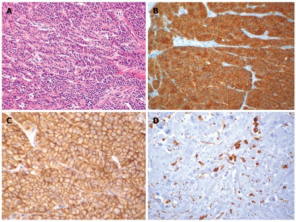
Figure 1 Histological images.
A: Lymph node metastasis of a high differentiated neuroendocrine carcinoma of the pancreas showing a trabecular pattern (HE, × 120); B The cells are positive for synaptophysin (× 120); C: High membranous expression of somatostatin-receptor SSTR-2 (× 120); D: Macrophages (CD68-positive) as a sign for tumor necrosis after PRRT (× 120).
- Citation: Kaemmerer D, Prasad V, Daffner W, Hörsch D, Klöppel G, Hommann M, Baum RP. Neoadjuvant peptide receptor radionuclide therapy for an inoperable neuroendocrine pancreatic tumor. World J Gastroenterol 2009; 15(46): 5867-5870
- URL: https://www.wjgnet.com/1007-9327/full/v15/i46/5867.htm
- DOI: https://dx.doi.org/10.3748/wjg.15.5867


