Copyright
©2009 The WJG Press and Baishideng.
World J Gastroenterol. Dec 14, 2009; 15(46): 5843-5850
Published online Dec 14, 2009. doi: 10.3748/wjg.15.5843
Published online Dec 14, 2009. doi: 10.3748/wjg.15.5843
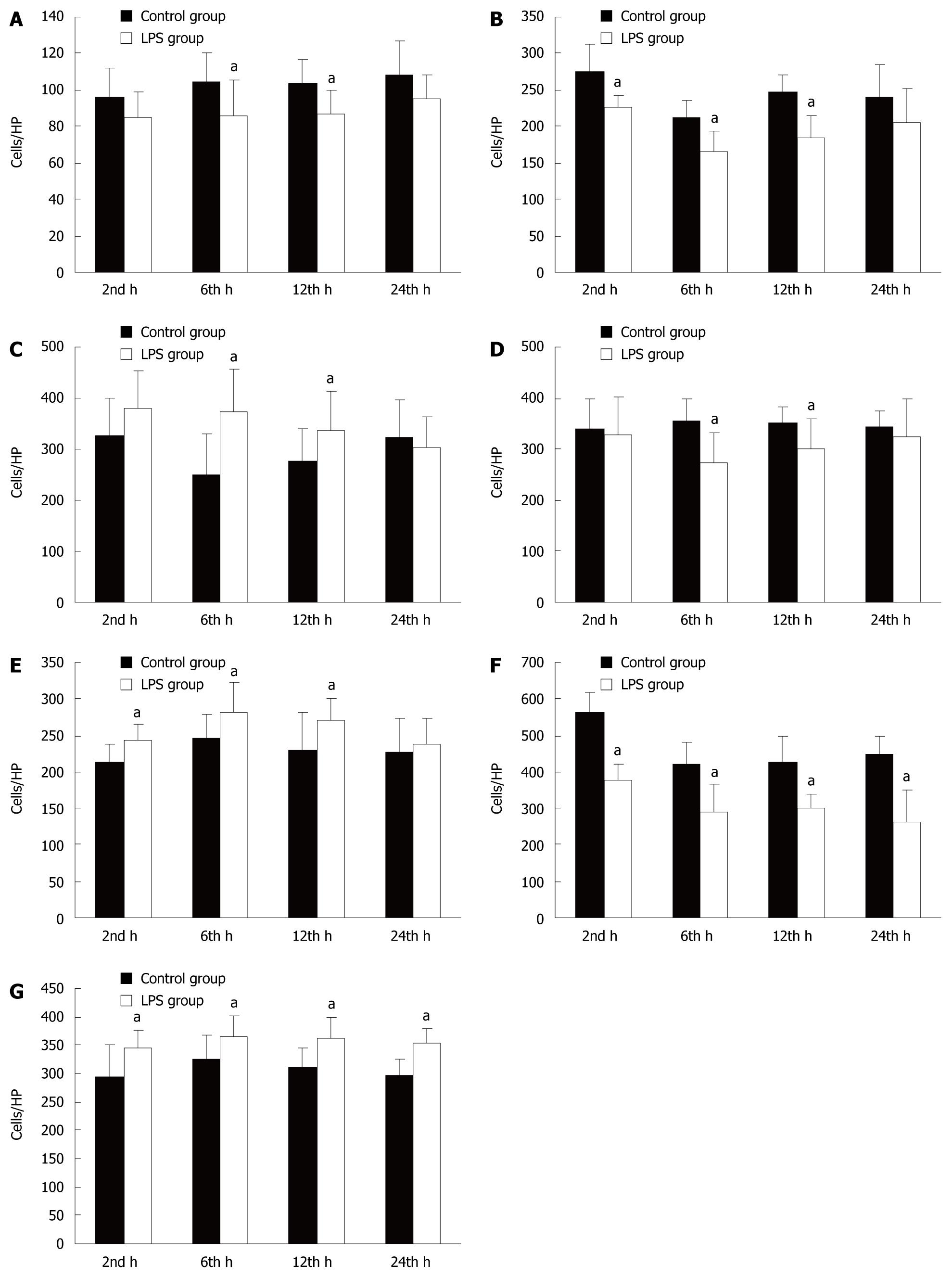
Figure 3 The number of immune cells and apoptotic lymphocytes in the intestinal mucosa.
A: The number of M-cells was significantly decreased after 6 and 12 h in the LPS group; B: The number of DCs was significantly decreased after 2, 6 and 12 h in the LPS group; C: The number of CD4+ T cells was significantly increased after 6 and 12 h before slightly decreasing by 24 h in the LPS group; D: The number of CD8+ T cells was significantly decreased after 6 and 12 h in the LPS group; E: The number of Tr cells was significantly increased after 2, 6 and 12 h in the LPS group; F: The number of IgA+ B cells was significantly increased at all time points in the LPS group; G: The number of apoptotic lymphocytes was significantly increased at all time points in the LPS group. aP < 0.05.
- Citation: Liu C, Li A, Weng YB, Duan ML, Wang BE, Zhang SW. Changes in intestinal mucosal immune barrier in rats with endotoxemia. World J Gastroenterol 2009; 15(46): 5843-5850
- URL: https://www.wjgnet.com/1007-9327/full/v15/i46/5843.htm
- DOI: https://dx.doi.org/10.3748/wjg.15.5843


