Copyright
©2009 The WJG Press and Baishideng.
World J Gastroenterol. Dec 14, 2009; 15(46): 5843-5850
Published online Dec 14, 2009. doi: 10.3748/wjg.15.5843
Published online Dec 14, 2009. doi: 10.3748/wjg.15.5843
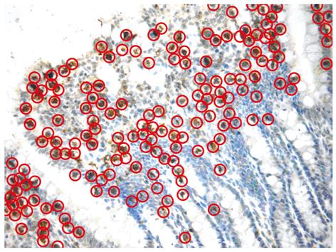
Figure 1 A sample image of immunohistochemical staining and TUNEL staining.
Some sample cells are circled to illustrate the positive cells that were counted (400 ×).
- Citation: Liu C, Li A, Weng YB, Duan ML, Wang BE, Zhang SW. Changes in intestinal mucosal immune barrier in rats with endotoxemia. World J Gastroenterol 2009; 15(46): 5843-5850
- URL: https://www.wjgnet.com/1007-9327/full/v15/i46/5843.htm
- DOI: https://dx.doi.org/10.3748/wjg.15.5843


