Copyright
©2009 The WJG Press and Baishideng.
World J Gastroenterol. Oct 21, 2009; 15(39): 4980-4983
Published online Oct 21, 2009. doi: 10.3748/wjg.15.4980
Published online Oct 21, 2009. doi: 10.3748/wjg.15.4980
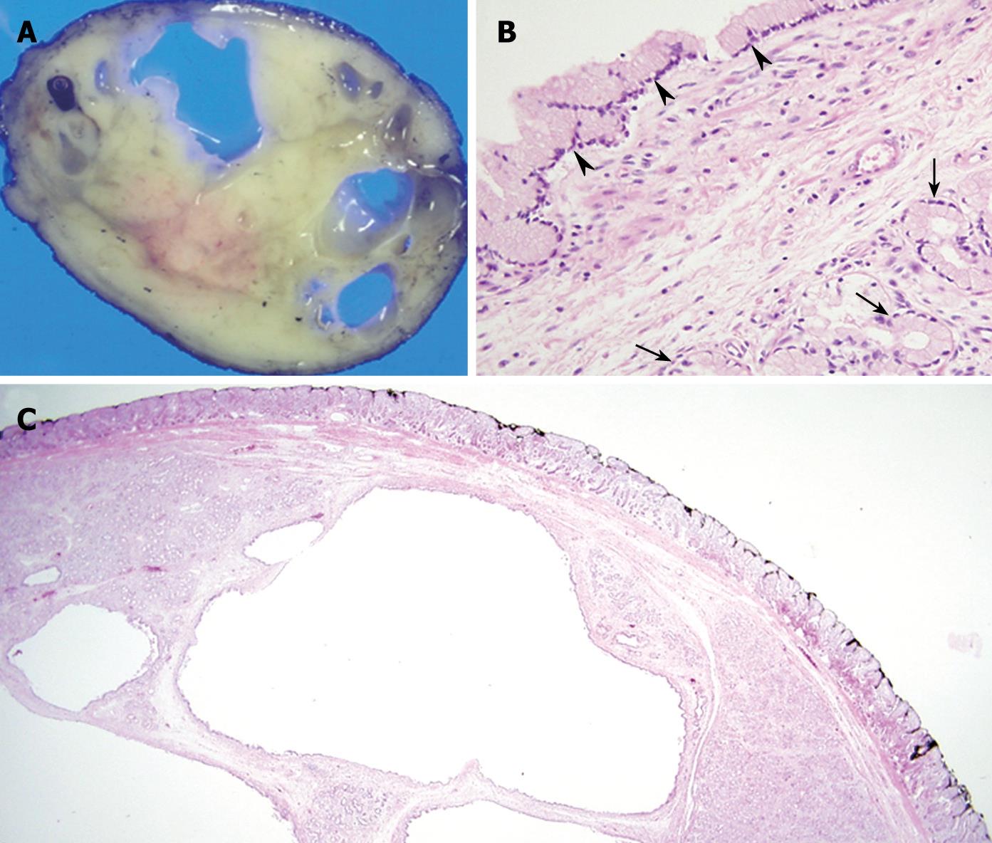
Figure 3 Histologic findings of the specimen.
A: Cut surface of the gross specimen showing a multiloculated cystic appearance filled with mucin-like material; B: Aggregated glands (arrows) are lined with cuboidal to columnar cells with abundant pale cytoplasm and a basally-located oval nucleus, resembling a normal Brunner’s gland in histopathologic findings of cystic Brunner’s gland hamartoma (HE, × 200) while the large cystic dilated gland showing the same lining (arrowheads) as a normal Brunner’s gland; C: Cystic Brunner’s gland hamartoma showing a well-demarcated nodular lesion, composed of a lobular collection of tubuloalveolar glands separated by fibrous septa, beneath the muscularis mucosa, in the low power field (× 12.5), while some glands showing cystic dilatation.
- Citation: Park BJ, Kim MJ, Lee JH, Park SS, Sung DJ, Cho SB. Cystic Brunner’s gland hamartoma in the duodenum: A case report. World J Gastroenterol 2009; 15(39): 4980-4983
- URL: https://www.wjgnet.com/1007-9327/full/v15/i39/4980.htm
- DOI: https://dx.doi.org/10.3748/wjg.15.4980


