Copyright
©2009 The WJG Press and Baishideng.
World J Gastroenterol. Aug 14, 2009; 15(30): 3757-3766
Published online Aug 14, 2009. doi: 10.3748/wjg.15.3757
Published online Aug 14, 2009. doi: 10.3748/wjg.15.3757
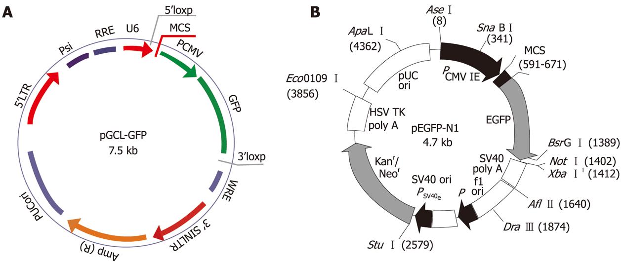
Figure 1 Structure of vectors.
A: Lentivirus vector pGCL-GFP containing a CMV driven GFP reporter and a U6 promoter upstream of cloning restriction sites (HpaI and XhoI) to allow the introduction of oligonucleotides encoding shRNAs. The multiple cloning site (MCS) is located between the U6 promoter and CMV. B: The pEGFP-N1 vector expressed green fluorescent protein following transfection into mammalian cells. MCS is located between the immediate early promoter of CMV and the EGFP. 1Methylated in the DNA provided by CLONTECH.
- Citation: Yang G, Huang C, Cao J, Huang KJ, Jiang T, Qiu ZJ. Lentivirus-mediated shRNA interference targeting STAT3 inhibits human pancreatic cancer cell invasion. World J Gastroenterol 2009; 15(30): 3757-3766
- URL: https://www.wjgnet.com/1007-9327/full/v15/i30/3757.htm
- DOI: https://dx.doi.org/10.3748/wjg.15.3757


