Copyright
©2009 The WJG Press and Baishideng.
World J Gastroenterol. Aug 7, 2009; 15(29): 3687-3690
Published online Aug 7, 2009. doi: 10.3748/wjg.15.3687
Published online Aug 7, 2009. doi: 10.3748/wjg.15.3687
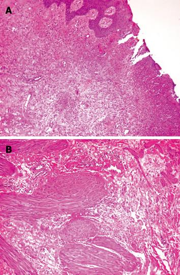
Figure 3 Histological features of the fibroepithelial polyp.
A: Photomicrograph of resected polyp showing fibrous tissue covered by squamous epithelium, small vessels and the ulceration of fibrous stroma (HE, × 100); B: Deeper layer of the polyp showing scattered smooth muscle fibers within the fibrous stroma (HE, × 100).
- Citation: Galanis I, Dragoumis D, Tsolakis M, Zarampoukas K, Zarampoukas T, Atmatzidis K. Obstructive ileus due to a giant fibroepithelial polyp of the anus. World J Gastroenterol 2009; 15(29): 3687-3690
- URL: https://www.wjgnet.com/1007-9327/full/v15/i29/3687.htm
- DOI: https://dx.doi.org/10.3748/wjg.15.3687


