Copyright
©2009 The WJG Press and Baishideng.
World J Gastroenterol. Aug 7, 2009; 15(29): 3670-3675
Published online Aug 7, 2009. doi: 10.3748/wjg.15.3670
Published online Aug 7, 2009. doi: 10.3748/wjg.15.3670
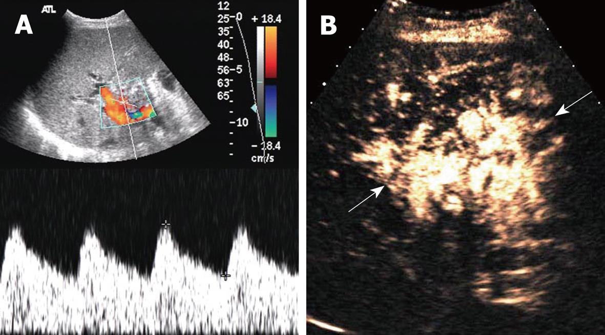
Figure 3 Hepatic artery obstruction with collateral circulation in a 36-year-old man who underwent RLDLT.
A: Longitudinal oblique Doppler US scan 6 mo after RLDLT revealed a tardus-parvus spectrum at the porta hepatis; B: Longitudinal oblique CEUS scan obtained following the Doppler scan. A reticulate vessel instead of the right hepatic artery trunk was seen (arrows) at the porta hepatis after ultrasound agent injection.
- Citation: Luo Y, Fan YT, Lu Q, Li B, Wen TF, Zhang ZW. CEUS: A new imaging approach for postoperative vascular complications after right-lobe LDLT. World J Gastroenterol 2009; 15(29): 3670-3675
- URL: https://www.wjgnet.com/1007-9327/full/v15/i29/3670.htm
- DOI: https://dx.doi.org/10.3748/wjg.15.3670


