Copyright
©2009 The WJG Press and Baishideng.
World J Gastroenterol. Jan 14, 2009; 15(2): 160-168
Published online Jan 14, 2009. doi: 10.3748/wjg.15.160
Published online Jan 14, 2009. doi: 10.3748/wjg.15.160
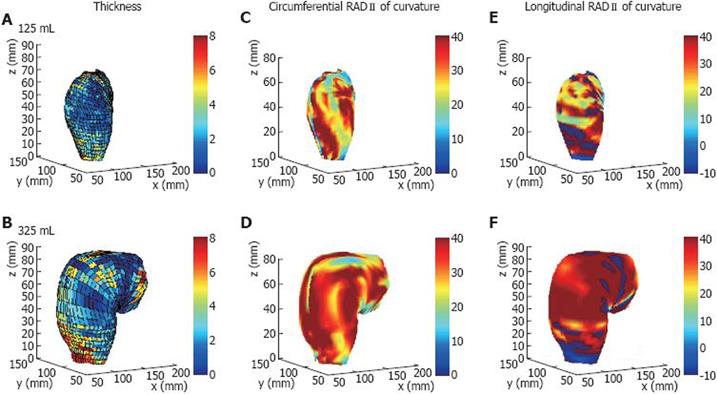
Figure 9 3D models of the rectum based on MRI and pressure recordings.
The 3D distribution of the rectal wall thickness (A-B), circumferential (C-D) and longitudinal (E-F) principal radii of curvatures in one healthy volunteer at infused volumes of 125 mL (A, C, E) and 325 mL (B, D, F). The change in colour from blue to red during bag distension indicates an increase in rectal wall thickness or radius of curvature, i.e. increase in diameter. Modified from [50].
- Citation: Frøkjær JB, Drewes AM, Gregersen H. Imaging of the gastrointestinal tract-novel technologies. World J Gastroenterol 2009; 15(2): 160-168
- URL: https://www.wjgnet.com/1007-9327/full/v15/i2/160.htm
- DOI: https://dx.doi.org/10.3748/wjg.15.160


