Copyright
©2009 The WJG Press and Baishideng.
World J Gastroenterol. Jan 14, 2009; 15(2): 160-168
Published online Jan 14, 2009. doi: 10.3748/wjg.15.160
Published online Jan 14, 2009. doi: 10.3748/wjg.15.160
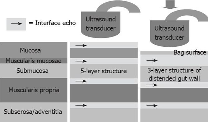
Figure 1 The principles of endosonography.
The histological gastrointestinal wall layers (left) are correlated to the typical layered ultrasound appearance of the gastrointestinal wall (middle). The 5-layered appearance is due to the addition of several interface echoes at the tissue interfaces. During compression or distension the wall is further stretched (including mucosal unfolding) which together usually obscures the second echo-rich mucosal layer. Hence, the wall appears 3-layered (see Figure 2).
- Citation: Frøkjær JB, Drewes AM, Gregersen H. Imaging of the gastrointestinal tract-novel technologies. World J Gastroenterol 2009; 15(2): 160-168
- URL: https://www.wjgnet.com/1007-9327/full/v15/i2/160.htm
- DOI: https://dx.doi.org/10.3748/wjg.15.160


