Copyright
©2008 The WJG Press and Baishideng.
World J Gastroenterol. Dec 28, 2008; 14(48): 7392-7396
Published online Dec 28, 2008. doi: 10.3748/wjg.14.7392
Published online Dec 28, 2008. doi: 10.3748/wjg.14.7392
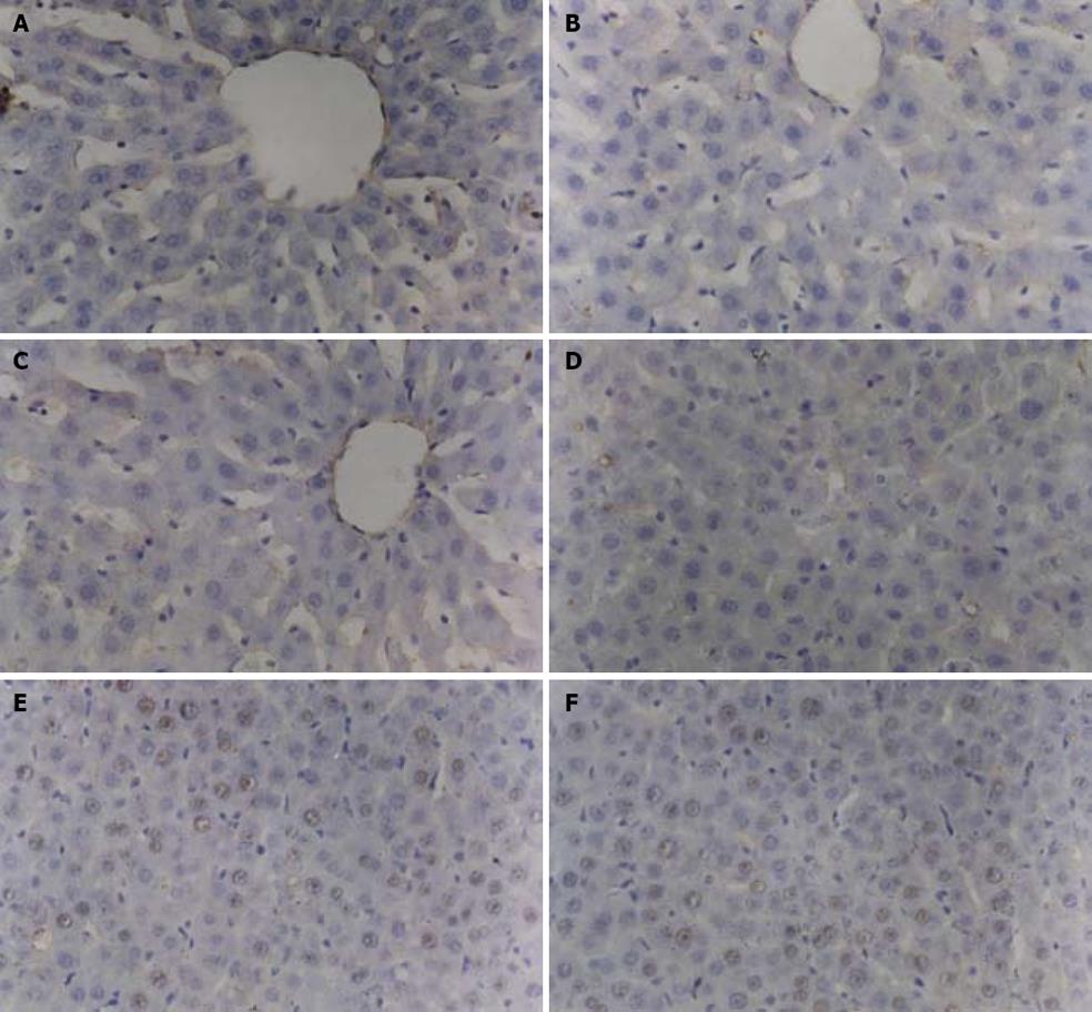
Figure 1 Positive cells are mainly hepatic parenchymal cells located near the sinus hepaticus and sinusoid endothelial cells of liver in melatonin exposure group (A, D), alcohol solvent control group (B, E), NS control group (C, F).
The positive cell rate of two control groups increased gradually and reached its peak 12 h after reperfusion, then decreased. This condition also occurred in melatonin exposure group, but the extent was much lower than that in the control groups.
- Citation: Li JY, Yin HZ, Gu X, Zhou Y, Zhang WH, Qin YM. Melatonin protects liver from intestine ischemia reperfusion injury in rats. World J Gastroenterol 2008; 14(48): 7392-7396
- URL: https://www.wjgnet.com/1007-9327/full/v14/i48/7392.htm
- DOI: https://dx.doi.org/10.3748/wjg.14.7392


