Copyright
©2008 The WJG Press and Baishideng.
World J Gastroenterol. Oct 14, 2008; 14(38): 5933-5937
Published online Oct 14, 2008. doi: 10.3748/wjg.14.5933
Published online Oct 14, 2008. doi: 10.3748/wjg.14.5933
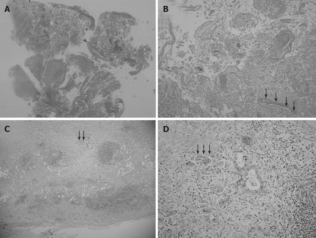
Figure 4 Low magnification view of the elevated lesion of the fundus showing obvious nodule necrosis.
(A, HE, loupe) including non-viable adenocarcinoma cells and remnants of glandular structures and vessel fragments (B, black arrow) (HE, × 40), viable cancer cell nests invading perimuscular connective tissue but not penetrating the serosa in the muscularis propria and subcutaneous layer beneath the necrotic nodule (C, black arrow) (HE, × 20), and a solid adenocarcinoma (D, black arrow) (HE, × 100).
- Citation: Hori T, Wagata T, Takemoto K, Shigeta T, Takuwa H, Hata K, Uemoto S, Yokoo N. Spontaneous necrosis of solid gallbladder adenocarcinoma accompanied with pancreaticobiliary maljunction. World J Gastroenterol 2008; 14(38): 5933-5937
- URL: https://www.wjgnet.com/1007-9327/full/v14/i38/5933.htm
- DOI: https://dx.doi.org/10.3748/wjg.14.5933


