Copyright
©2008 The WJG Press and Baishideng.
World J Gastroenterol. Oct 14, 2008; 14(38): 5933-5937
Published online Oct 14, 2008. doi: 10.3748/wjg.14.5933
Published online Oct 14, 2008. doi: 10.3748/wjg.14.5933
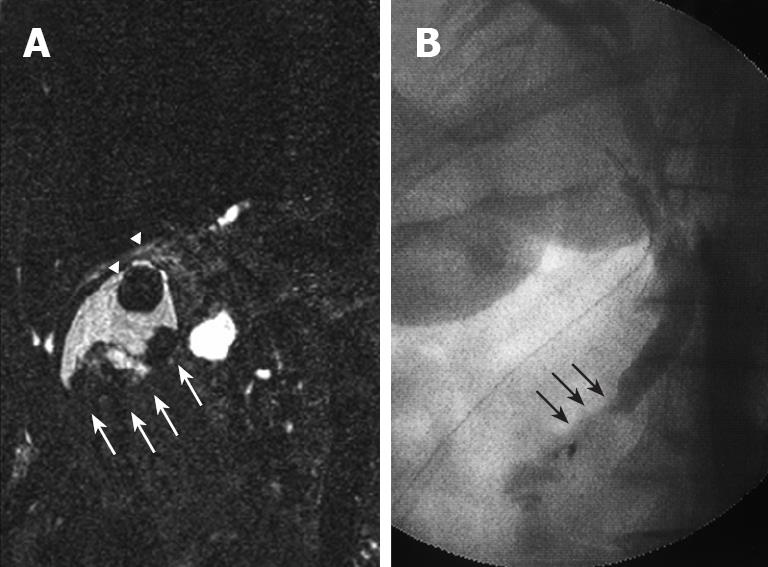
Figure 3 MRC.
(A) showing irregular defects due to the elevated lesion of the fundus (white arrow) and a round defect due to stone incarceration in the neck (white arrow head), intra-operative cholangiography (B) revealing PBM and a 15 mm long common channel (black arrow).
- Citation: Hori T, Wagata T, Takemoto K, Shigeta T, Takuwa H, Hata K, Uemoto S, Yokoo N. Spontaneous necrosis of solid gallbladder adenocarcinoma accompanied with pancreaticobiliary maljunction. World J Gastroenterol 2008; 14(38): 5933-5937
- URL: https://www.wjgnet.com/1007-9327/full/v14/i38/5933.htm
- DOI: https://dx.doi.org/10.3748/wjg.14.5933


