Copyright
©2008 The WJG Press and Baishideng.
World J Gastroenterol. Oct 14, 2008; 14(38): 5933-5937
Published online Oct 14, 2008. doi: 10.3748/wjg.14.5933
Published online Oct 14, 2008. doi: 10.3748/wjg.14.5933
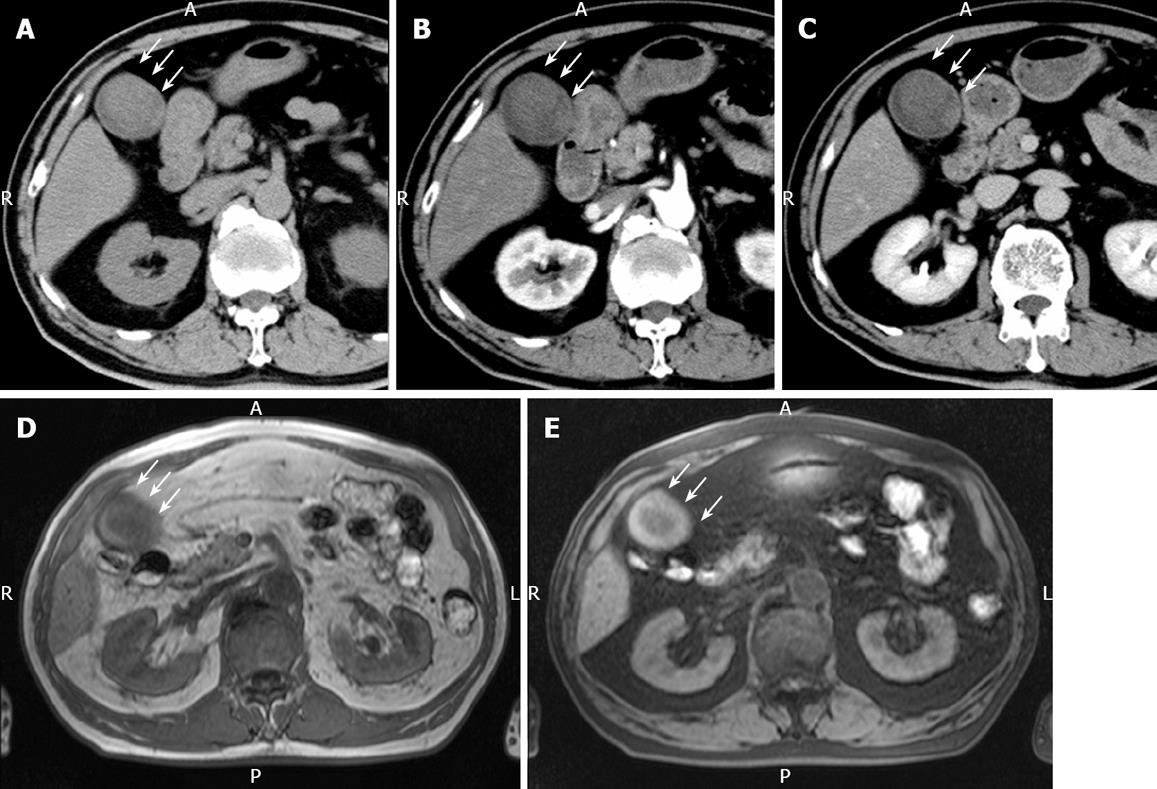
Figure 2 Plain CT.
(A) showing a relatively low-density mass of the fundus measuring 5 cm x 4 cm in diameter, contrast-enhanced CT revealing no positive enhancement of this mass-like lesion in its early (B) and late (C) phases, MRI showing a mass in the fundus with a slightly low intensity on T1-weighted images (D) and a slightly high intensity on T2-weighted images (E). White arrow represents the mass-like lesion of the fundus.
- Citation: Hori T, Wagata T, Takemoto K, Shigeta T, Takuwa H, Hata K, Uemoto S, Yokoo N. Spontaneous necrosis of solid gallbladder adenocarcinoma accompanied with pancreaticobiliary maljunction. World J Gastroenterol 2008; 14(38): 5933-5937
- URL: https://www.wjgnet.com/1007-9327/full/v14/i38/5933.htm
- DOI: https://dx.doi.org/10.3748/wjg.14.5933


