Copyright
©2008 The WJG Press and Baishideng.
World J Gastroenterol. Oct 14, 2008; 14(38): 5933-5937
Published online Oct 14, 2008. doi: 10.3748/wjg.14.5933
Published online Oct 14, 2008. doi: 10.3748/wjg.14.5933
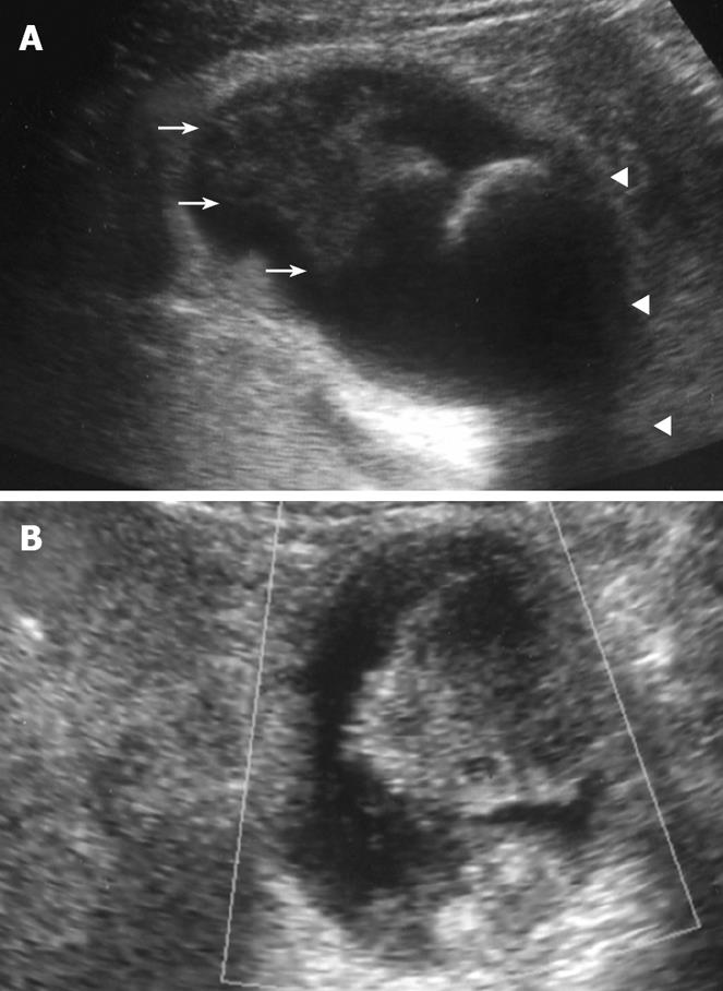
Figure 1 Plain US.
(A) of the GB showing wall thickening of the neck and body (consistent with cholecystitis), a strong echoic level with acoustic shadow at the neck (consistent with stones), and a mass-like lesion of the fundus, and Doppler US (B) revealing no blood flow in the mass-like lesion.
- Citation: Hori T, Wagata T, Takemoto K, Shigeta T, Takuwa H, Hata K, Uemoto S, Yokoo N. Spontaneous necrosis of solid gallbladder adenocarcinoma accompanied with pancreaticobiliary maljunction. World J Gastroenterol 2008; 14(38): 5933-5937
- URL: https://www.wjgnet.com/1007-9327/full/v14/i38/5933.htm
- DOI: https://dx.doi.org/10.3748/wjg.14.5933


