Copyright
©2008 The WJG Press and Baishideng.
World J Gastroenterol. Sep 14, 2008; 14(34): 5290-5300
Published online Sep 14, 2008. doi: 10.3748/wjg.14.5290
Published online Sep 14, 2008. doi: 10.3748/wjg.14.5290
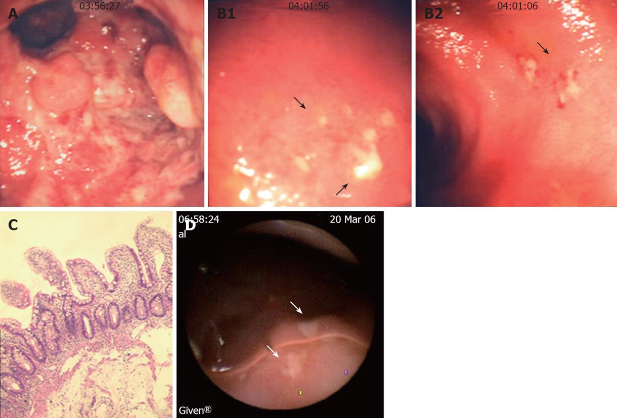
Figure 4 Endoscopic view, histological analysis and WCE images of the rectum and neo-terminal ileum from a third UC patient (LRA) with IRA.
A: Endoscopic view of the rectal stump showing erosions and pseudopolyps; B: Endoscopic views of the ileum showing small focal ulcers above the anastomosis; C: Ileal biopsy sample from the same patient showing no villous atrophy or colonic metaplasia by histology; D: WCE images showing in the neo-terminal ileum above the anastomosis, 2 ulcers covered by fibrin (arrows) surrounded by macroscopically normal ileum.
- Citation: Biancone L, Calabrese E, Palmieri G, Petruzziello C, Onali S, Sica GS, Cossignani M, Condino G, Das KM, Pallone F. Ileal lesions in patients with ulcerative colitis after ileo-rectal anastomosis: Relationship with colonic metaplasia. World J Gastroenterol 2008; 14(34): 5290-5300
- URL: https://www.wjgnet.com/1007-9327/full/v14/i34/5290.htm
- DOI: https://dx.doi.org/10.3748/wjg.14.5290


