Copyright
©2008 The WJG Press and Baishideng.
World J Gastroenterol. Aug 14, 2008; 14(30): 4834-4837
Published online Aug 14, 2008. doi: 10.3748/wjg.14.4834
Published online Aug 14, 2008. doi: 10.3748/wjg.14.4834
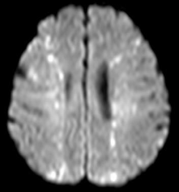
Figure 4 Diffusion-weighted MR images obtained 48 h after TACE shows multiple hyperattenuating and hypertensive punctate-patchy lesions in both cerebral hemispheres.
- Citation: Choi CS, Kim KH, Seo GS, Cho EY, Oh HJ, Choi SC, Kim TH, Kim HC, Roh BS. Cerebral and pulmonary embolisms after transcatheter arterial chemoembolization for hepatocellular carcinoma. World J Gastroenterol 2008; 14(30): 4834-4837
- URL: https://www.wjgnet.com/1007-9327/full/v14/i30/4834.htm
- DOI: https://dx.doi.org/10.3748/wjg.14.4834


