Copyright
©2008 The WJG Press and Baishideng.
World J Gastroenterol. Jul 14, 2008; 14(26): 4257-4259
Published online Jul 14, 2008. doi: 10.3748/wjg.14.4257
Published online Jul 14, 2008. doi: 10.3748/wjg.14.4257
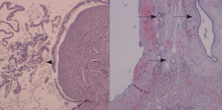
Figure 4 The wall of the largest cyst showing papillary projections of the lining epithelium (arrowhead), presence of microscopic cysts with the same epithelial lining (arrows), calcifications and hyaline degeneration of the stroma (HE, × 40).
- Citation: Manouras A, Lagoudianakis E, Alevizos L, Markogiannakis H, Kafiri G, Bramis C, Filis K, Toutouzas K. Laparoscopic fenestration of multiple giant biliary mucinous cystadenomas of the liver. World J Gastroenterol 2008; 14(26): 4257-4259
- URL: https://www.wjgnet.com/1007-9327/full/v14/i26/4257.htm
- DOI: https://dx.doi.org/10.3748/wjg.14.4257


