Copyright
©2008 The WJG Press and Baishideng.
World J Gastroenterol. May 14, 2008; 14(18): 2935-2938
Published online May 14, 2008. doi: 10.3748/wjg.14.2935
Published online May 14, 2008. doi: 10.3748/wjg.14.2935
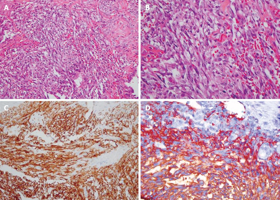
Figure 3 Pathologic findings.
A and B: Atypical cells forming solid sheets filled with RBCs (HE stain, × 200, × 400, respectively); C and D: Immunohistochemical staining for CD31 and CD34 showing diffuse strong positive expression (× 200, × 400, respectively).
- Citation: Lee SW, Song CY, Gi YH, Kang SB, Kim YS, Nam SW, Lee DS, Kim JO. Hepatic angiosarcoma manifested as recurrent hemoperitoneum. World J Gastroenterol 2008; 14(18): 2935-2938
- URL: https://www.wjgnet.com/1007-9327/full/v14/i18/2935.htm
- DOI: https://dx.doi.org/10.3748/wjg.14.2935


