Copyright
©2008 The WJG Press and Baishideng.
World J Gastroenterol. May 14, 2008; 14(18): 2935-2938
Published online May 14, 2008. doi: 10.3748/wjg.14.2935
Published online May 14, 2008. doi: 10.3748/wjg.14.2935
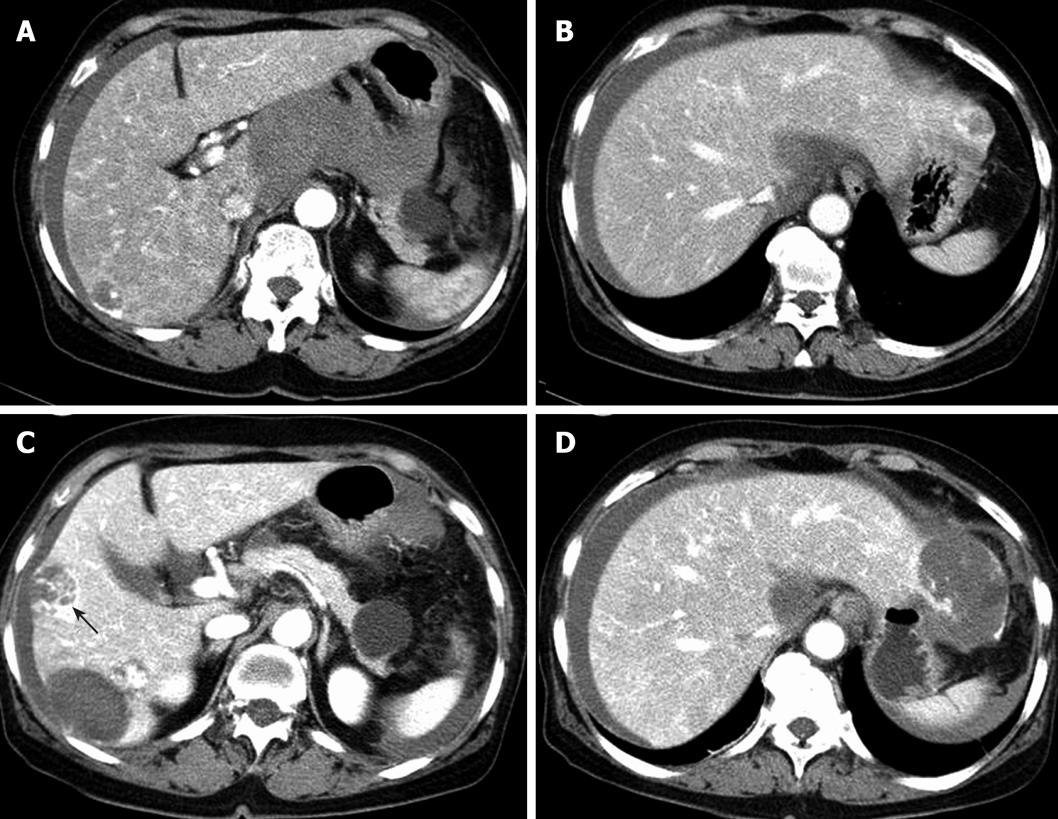
Figure 1 Initial abdominal computed tomography images.
A and B: A large amount of hematoma in lesser sac and a lowly attenuated nodular lesion with peripheral globular enhancement in the left lobe. Abdominal CT images after 1 mo; C and D: increased size of lowly attenuated lesions in the right lobe and the left lateral lobe. One showing septum-like appearance and peripheral enhancement within the mass (black arrow).
- Citation: Lee SW, Song CY, Gi YH, Kang SB, Kim YS, Nam SW, Lee DS, Kim JO. Hepatic angiosarcoma manifested as recurrent hemoperitoneum. World J Gastroenterol 2008; 14(18): 2935-2938
- URL: https://www.wjgnet.com/1007-9327/full/v14/i18/2935.htm
- DOI: https://dx.doi.org/10.3748/wjg.14.2935


