Copyright
©2007 Baishideng Publishing Group Co.
World J Gastroenterol. Feb 28, 2007; 13(8): 1252-1256
Published online Feb 28, 2007. doi: 10.3748/wjg.v13.i8.1252
Published online Feb 28, 2007. doi: 10.3748/wjg.v13.i8.1252
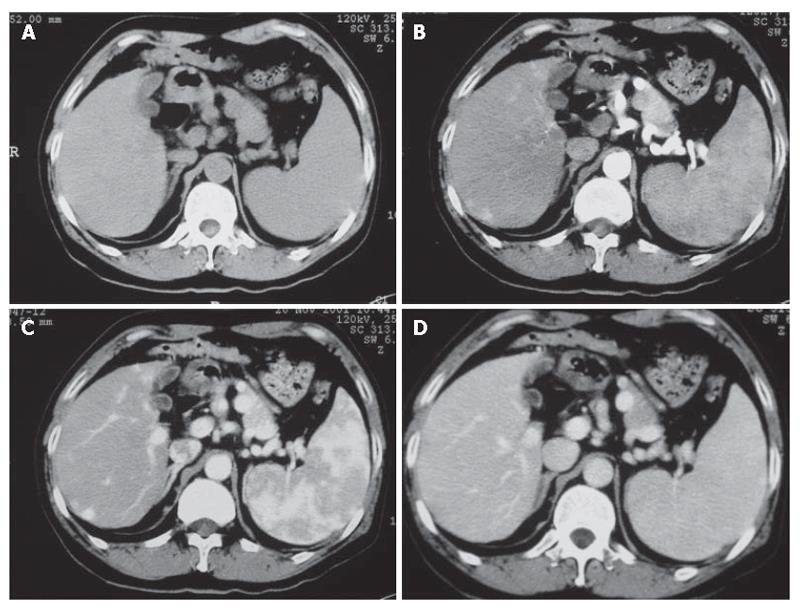
Figure 1 A 42-yr old man with pathologically proven he-patocellular carcinoma.
A: Plan scanning; B: early arterial phase MDCT image showing slightly enhanced nodule in right lobe; C: later arterial phase MDCT image showing obvious enhanced lesion; D: on the portal venous phase MDCT image showing slight low attenuation.
- Citation: Zhao H, Yao JL, Wang Y, Zhou KR. Detection of small hepatocellular carcinoma: Comparison of dynamic enhancement magnetic resonance imaging and multiphase multirow-detector helical CT scanning. World J Gastroenterol 2007; 13(8): 1252-1256
- URL: https://www.wjgnet.com/1007-9327/full/v13/i8/1252.htm
- DOI: https://dx.doi.org/10.3748/wjg.v13.i8.1252


