Copyright
©2007 Baishideng Publishing Group Co.
World J Gastroenterol. Feb 14, 2007; 13(6): 858-865
Published online Feb 14, 2007. doi: 10.3748/wjg.v13.i6.858
Published online Feb 14, 2007. doi: 10.3748/wjg.v13.i6.858
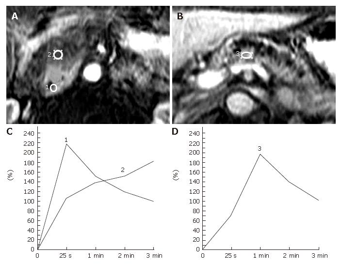
Figure 2 Representative pancreatic TIC profiles in patients with pancreatic ductal carcinoma developed in a normal pancreas.
A, B: Dynamic contrast-enhanced MRI images of the pancreas in a 59-year-old man with carcinoma of the head of the pancreas. The ROIs are placed at the pancreatic mass (No.2 ROI) and the non-tumorous pancreatic parenchyma both proximal (No.1 ROI) and distal (No.3 ROI) to the mass lesion; C: Pancreatic TICs obtained from the no.1 and no. 2 ROIs as in (A) demonstrate type-I and type-IV, respectively; D: Pancreatic TIC obtained from the No.3 ROI as in Figure 2B shows type-II.
- Citation: Tajima Y, Kuroki T, Tsutsumi R, Isomoto I, Uetani M, Kanematsu T. Pancreatic carcinoma coexisting with chronic pancreatitis versus tumor-forming pancreatitis: Diagnostic utility of the time-signal intensity curve from dynamic contrast-enhanced MR imaging. World J Gastroenterol 2007; 13(6): 858-865
- URL: https://www.wjgnet.com/1007-9327/full/v13/i6/858.htm
- DOI: https://dx.doi.org/10.3748/wjg.v13.i6.858


