Copyright
©2007 Baishideng Publishing Group Inc.
World J Gastroenterol. Nov 21, 2007; 13(43): 5771-5774
Published online Nov 21, 2007. doi: 10.3748/wjg.v13.i43.5771
Published online Nov 21, 2007. doi: 10.3748/wjg.v13.i43.5771
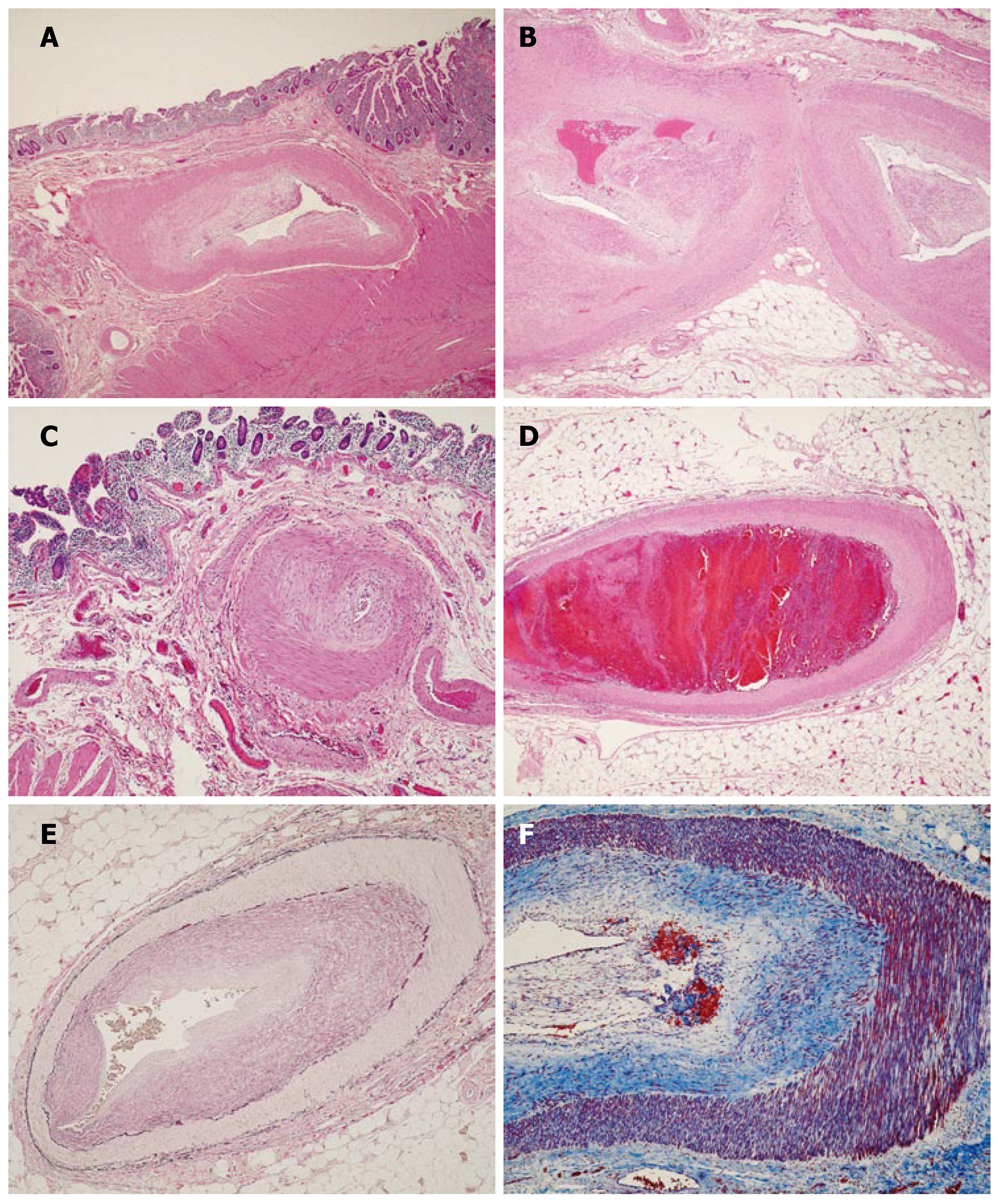
Figure 1 Microscopic sections showing marked thickening (A) and hyalinization (B) of medium sized vessel walls with prominent eccentric intimal proliferation with no arteritis, necrosis, inflammation and calcification (hematoxylin-eosin, original magnification × 200); sparing of small sized vessels (C) (hematoxylin-eosin, original magnification × 200); focal evidence of recent vascular thrombosis with early organization in one vessel (D) (hematoxylin-eosin, original magnification × 200); Verhoeff’s Van Gieson elastic stain highlighting the prominent intimal hyperplasia (E) (original magnification × 200; Masson’s trichrome stain highlighting a loose matrix of fibrous tissue replacing and expanding the intima (F) (original magnification × 200).
- Citation: Rodriguez Urrego PA, Flanagan M, Tsai WS, Rezac C, Barnard N. Massive gastrointestinal bleeding: An unusual case of asymptomatic extrarenal, visceral, fibromuscular dysplasia. World J Gastroenterol 2007; 13(43): 5771-5774
- URL: https://www.wjgnet.com/1007-9327/full/v13/i43/5771.htm
- DOI: https://dx.doi.org/10.3748/wjg.v13.i43.5771


