Copyright
©2007 Baishideng Publishing Group Inc.
World J Gastroenterol. Nov 14, 2007; 13(42): 5664-5667
Published online Nov 14, 2007. doi: 10.3748/wjg.v13.i42.5664
Published online Nov 14, 2007. doi: 10.3748/wjg.v13.i42.5664
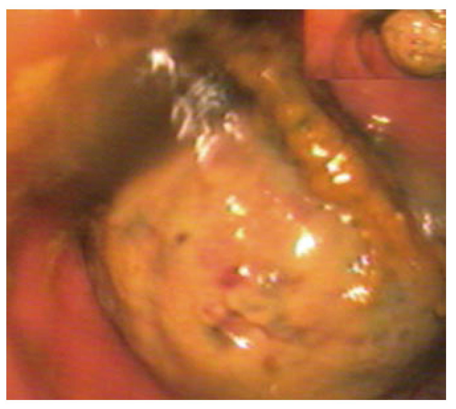
Figure 1 Colonoscopy showed a yellowish, spherical, polypoid lesion, with a lot of inflammatory necrotic tissue and numerous areas of ulceration on its surface, which arose from the lateral wall of the colon.
The lesion almost obstructed the whole lumen.
- Citation: Jiang L, Jiang LS, Li FY, Ye H, Li N, Cheng NS, Zhou Y. Giant submucosal lipoma located in the descending colon: A case report and review of the literature. World J Gastroenterol 2007; 13(42): 5664-5667
- URL: https://www.wjgnet.com/1007-9327/full/v13/i42/5664.htm
- DOI: https://dx.doi.org/10.3748/wjg.v13.i42.5664


