Copyright
©2007 Baishideng Publishing Group Co.
World J Gastroenterol. Jan 28, 2007; 13(4): 532-537
Published online Jan 28, 2007. doi: 10.3748/wjg.v13.i4.532
Published online Jan 28, 2007. doi: 10.3748/wjg.v13.i4.532
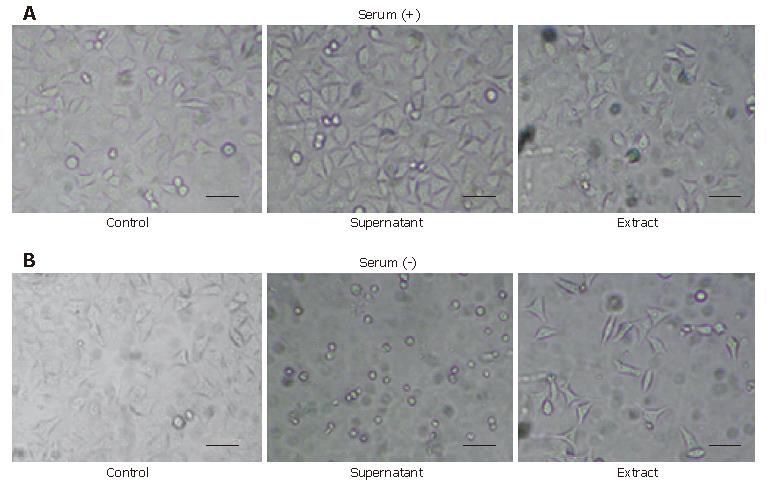
Figure 1 Morphologic change of HeLa cells caused by the supernatant and the extract of H pylori with or without serum after 24 h.
HeLa cells were seeded at 5 000 cells per well in every plate. The concentration of the supernatant cultured with or without serum was 2.1 mg/mL and the concentration of the respective extract was 0.5 mg/mL. A: HeLa cells exposed to the supernatant of the method using serum showed little change and the ones exposed to the extract also showed no remarkable changes but included a few minor vacuolations compared to the control; B: HeLa cells exposed to the supernatant of the method without serum showed a round-formed shape and a decayed contour with greatly decreased cell number, and those exposed to its extract showed an elongated or spindle-like shape with a decrease in cell number. Scale bar, 200 μm.
-
Citation: Ohno H, Murano A. Serum-free culture of
H pylori intensifies cytotoxicity. World J Gastroenterol 2007; 13(4): 532-537 - URL: https://www.wjgnet.com/1007-9327/full/v13/i4/532.htm
- DOI: https://dx.doi.org/10.3748/wjg.v13.i4.532


