Copyright
©2007 Baishideng Publishing Group Inc.
World J Gastroenterol. Oct 21, 2007; 13(39): 5226-5231
Published online Oct 21, 2007. doi: 10.3748/wjg.v13.i39.5226
Published online Oct 21, 2007. doi: 10.3748/wjg.v13.i39.5226
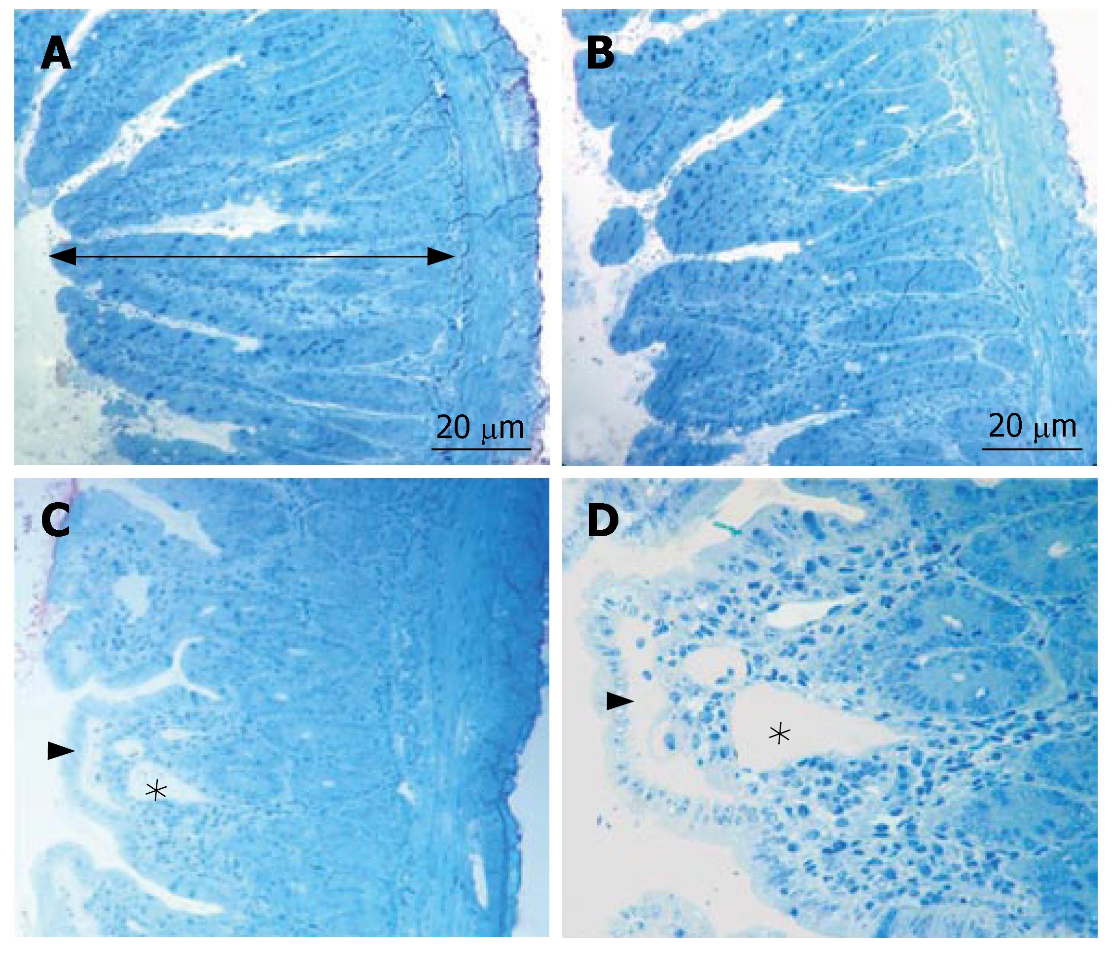
Figure 1 The micrographs of light microscope stained with toluidin blue.
A: Typical structure of villi and the total mucosal thickness (arrow) in the groupI; B: The normal villous architecture in group III; C, D: Blunting of the villi, the subepithelial edema (arrow head) and the dilated the lacteal (*) in group II.
- Citation: Sabuncuoglu MZ, Kismet K, Kilicoglu SS, Kilicoglu B, Erel S, Muratoglu S, Sunay AE, Erdemli E, Akkus MA. Propolis reduces bacterial translocation and intestinal villus atrophy in experimental obstructive jaundice. World J Gastroenterol 2007; 13(39): 5226-5231
- URL: https://www.wjgnet.com/1007-9327/full/v13/i39/5226.htm
- DOI: https://dx.doi.org/10.3748/wjg.v13.i39.5226


