Copyright
©2007 Baishideng Publishing Group Co.
World J Gastroenterol. Sep 21, 2007; 13(35): 4746-4754
Published online Sep 21, 2007. doi: 10.3748/wjg.v13.i35.4746
Published online Sep 21, 2007. doi: 10.3748/wjg.v13.i35.4746
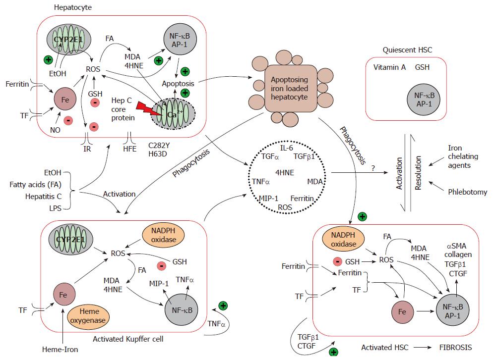
Figure 2 Mechanisms of iron-induced liver injury and fibrosis.
Iron catalyses the formation of several reactive oxygen species in hepatocytes. Under normal circumstances, hepatocytes are able to effectively cope with oxidant stress. When the liver is subjected to a secondary insult that enhances hepatic oxidant stress, hepatic fibrosis begins to develop. Increased oxidative stress and other pathological modes of action of HCV, ethanol and steatosis, lead to mitochondrial dysfunction and hepatocyte apoptosis. Kupffer cell activation is achieved by phagocytosis of apoptosing hepatocytes in conjunction with the direct effects of iron, HCV infection, ethanol and steatosis on Kupffer cells. The concomitant hepatocyte damage/apoptosis and Kupffer cell activation is able to drive and maintain hepatic stellate cell activation leading to fibrosis and ultimately cirrhosis if left unchecked. Fe, iron; TF, transferrin; ROS, reactive oxygen species; FA, fatty acids; IR, insulin receptor; GSH, glutathione; TNFα, tumour necrosis factor α; NO, nitric oxide; IL-6, interleukin 6; TGFβ1, transforming growth factor β1; CTGF, connective tissue growth factor; αSMA, α smooth muscle actin; MDA, malondialdehyde; 4HNE, 4-hydroxynonenal; MIP-1, macrophage inflammatory protein-1; TGFα, transforming growth factor α; NF-κB, nuclear factor κB; AP-1, activating protein-1.
- Citation: Philippe MA, Ruddell RG, Ramm GA. Role of iron in hepatic fibrosis: One piece in the puzzle. World J Gastroenterol 2007; 13(35): 4746-4754
- URL: https://www.wjgnet.com/1007-9327/full/v13/i35/4746.htm
- DOI: https://dx.doi.org/10.3748/wjg.v13.i35.4746


