Copyright
©2007 Baishideng Publishing Group Co.
World J Gastroenterol. Sep 14, 2007; 13(34): 4649-4652
Published online Sep 14, 2007. doi: 10.3748/wjg.v13.i34.4649
Published online Sep 14, 2007. doi: 10.3748/wjg.v13.i34.4649
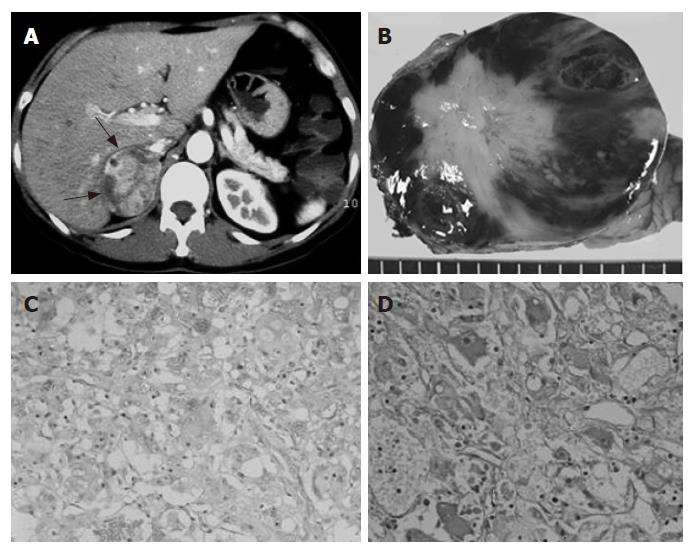
Figure 1 A: Enhanced abdominal CT scan showing a right adrenal tumor (arrows); B: Cut section of the tumor showing variegated white and red-brown areas; C: Immunohistochemical detection of chromogranin A in the tumor cells (× 200); D: Immunohistochemical detection of VIP-positive cells.
Stained cells were scattered throughout the tumor tissue (× 300).
- Citation: Ikuta SI, Yasui C, Kawanaka M, Aihara T, Yoshie H, Yanagi H, Mitsunobu M, Sugihara A, Yamanaka N. Watery diarrhea, hypokalemia and achlorhydria syndrome due to an adrenal pheochromocytoma. World J Gastroenterol 2007; 13(34): 4649-4652
- URL: https://www.wjgnet.com/1007-9327/full/v13/i34/4649.htm
- DOI: https://dx.doi.org/10.3748/wjg.v13.i34.4649


