Copyright
©2007 Baishideng Publishing Group Co.
World J Gastroenterol. Jan 7, 2007; 13(1): 65-73
Published online Jan 7, 2007. doi: 10.3748/wjg.v13.i1.65
Published online Jan 7, 2007. doi: 10.3748/wjg.v13.i1.65
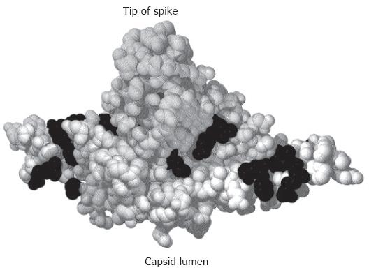
Figure 2 Crystal structure of a C-terminally truncated C protein dimer[21].
The spike protrudes upwards. The lumen of the capsid would be below the figure. Mutational analysis identified aa (shown in black) where the mutation was compatible with capsid formation and viral DNA synthesis in the lumen of the particle but blocked nucleocapsid envelopment[121].
- Citation: Bruss V. Hepatitis B virus morphogenesis. World J Gastroenterol 2007; 13(1): 65-73
- URL: https://www.wjgnet.com/1007-9327/full/v13/i1/65.htm
- DOI: https://dx.doi.org/10.3748/wjg.v13.i1.65


