Copyright
©2006 Baishideng Publishing Group Co.
World J Gastroenterol. Mar 7, 2006; 12(9): 1439-1442
Published online Mar 7, 2006. doi: 10.3748/wjg.v12.i9.1439
Published online Mar 7, 2006. doi: 10.3748/wjg.v12.i9.1439
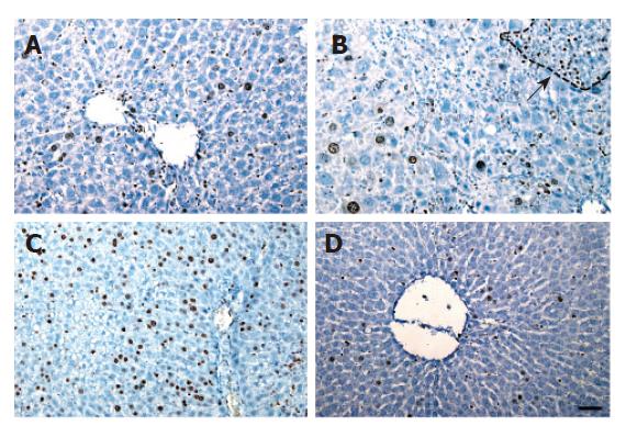
Figure 3 Ki-67 immunohistochemical analysis of rat liver.
A: A few hepatocytes were Ki-67 positive. B: Ki-67 positive hepatocytes were mainly found in small hepatocyte nodules as the arrow indicated. C: Abundant Ki-67 positive cells. D: Only a few hepatocytes were Ki-67 positive in rat liver.
- Citation: Zhou XF, Wang Q, Chu JX, Liu AL. Effects of retrorsine on mouse hepatocyte proliferation after liver injury. World J Gastroenterol 2006; 12(9): 1439-1442
- URL: https://www.wjgnet.com/1007-9327/full/v12/i9/1439.htm
- DOI: https://dx.doi.org/10.3748/wjg.v12.i9.1439


