Copyright
©2006 Baishideng Publishing Group Co.
World J Gastroenterol. Feb 21, 2006; 12(7): 1153-1156
Published online Feb 21, 2006. doi: 10.3748/wjg.v12.i7.1153
Published online Feb 21, 2006. doi: 10.3748/wjg.v12.i7.1153
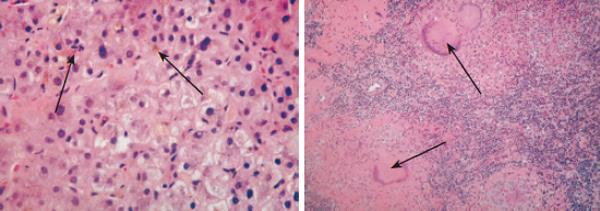
Figure 3 Hematoxylin and eosin (H and E) staining of liver tissue (A) and mediastinal lymph node (B) showing bile pigments (thick arrow) with bile duct proliferation (thin arrow) and inflammatory cell infiltrate and epethelioid granuloma with many langerhans cells but no caseation (arrows).
- Citation: Alsawat KE, Aljebreen AM. Resolution of tuberculous biliary stricture after medical therapy. World J Gastroenterol 2006; 12(7): 1153-1156
- URL: https://www.wjgnet.com/1007-9327/full/v12/i7/1153.htm
- DOI: https://dx.doi.org/10.3748/wjg.v12.i7.1153


