Copyright
©2006 Baishideng Publishing Group Co.
World J Gastroenterol. Feb 7, 2006; 12(5): 800-803
Published online Feb 7, 2006. doi: 10.3748/wjg.v12.i5.800
Published online Feb 7, 2006. doi: 10.3748/wjg.v12.i5.800
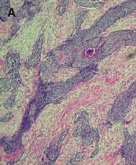
Figure 4 A: Photomicrography depicts nests or clusters of small tumor cells outlined by characteristic desmoplastic stroma bands (hematoxylin and eosin staining, 100×).
B: Photomicrography demonstrates the monomorphic, small round tumor cell population with a small spherical or spheroid nucleus, rich in chromatin (hematoxylin and eosin staining, 400×). C: The immunohistochemical staining of tumor cells is positive for desmin.
- Citation: Chang CC, Hsu JT, Tseng JH, Hwang TL, Chen HM, Jan YY. Combined resection and multi-agent adjuvant chemotherapy for desmoplastic small round cell tumor arising in the abdominal cavity: Report of a case. World J Gastroenterol 2006; 12(5): 800-803
- URL: https://www.wjgnet.com/1007-9327/full/v12/i5/800.htm
- DOI: https://dx.doi.org/10.3748/wjg.v12.i5.800


