Copyright
©2006 Baishideng Publishing Group Co.
World J Gastroenterol. Dec 7, 2006; 12(45): 7380-7387
Published online Dec 7, 2006. doi: 10.3748/wjg.v12.i45.7380
Published online Dec 7, 2006. doi: 10.3748/wjg.v12.i45.7380
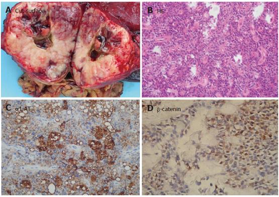
Figure 4 Solid pseudo-papillary neoplasia.
A: Cystic structures containing hemorrhagic debris surrounded by hemorrhagic-necrotic tissue; B: SPN showing solid areas consisting of monotonous polygonal epithelioid cells, often with minimal intervening stroma accompanied with innumerable capillary-sized vessels. In the pseudopapillary regions, the cells away from the small vessels appeared to have dropped away, leaving an irregular cuff of cells surrounding each vascular core (HE × 100); C: SPN showing a consistent pattern of reactivity for α-1-AT (Envison × 100); D: Positive β-catenin and progesteron receptor limited to the nuclei of tumor cells (Envison × 200).
- Citation: Ji Y, Lou WH, Jin DY, Kuang TT, Zeng MS, Tan YS, Zeng HY, Sujie A, Zhu XZ. A series of 64 cases of pancreatic cystic neoplasia from an institutional study of China. World J Gastroenterol 2006; 12(45): 7380-7387
- URL: https://www.wjgnet.com/1007-9327/full/v12/i45/7380.htm
- DOI: https://dx.doi.org/10.3748/wjg.v12.i45.7380


