Copyright
©2006 Baishideng Publishing Group Co.
World J Gastroenterol. Dec 7, 2006; 12(45): 7380-7387
Published online Dec 7, 2006. doi: 10.3748/wjg.v12.i45.7380
Published online Dec 7, 2006. doi: 10.3748/wjg.v12.i45.7380
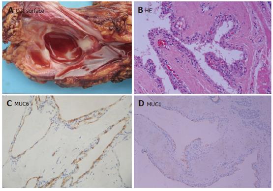
Figure 2 Serous cystic neoplasm.
A: Smooth and thin cyst wall on cut surface containing clear red-brown fulid; B: Cysts wall lined by a single layer of cuboidal or flattened epithelial cells with pale to clear cytoplasm, and centrally located tumor cells with round to oval nuclei (HE × 100); C: Tumor cells strongly positive for MUC6 (Envison × 100); D: MUC1 expression in some tumor cells (Envison × 40).
- Citation: Ji Y, Lou WH, Jin DY, Kuang TT, Zeng MS, Tan YS, Zeng HY, Sujie A, Zhu XZ. A series of 64 cases of pancreatic cystic neoplasia from an institutional study of China. World J Gastroenterol 2006; 12(45): 7380-7387
- URL: https://www.wjgnet.com/1007-9327/full/v12/i45/7380.htm
- DOI: https://dx.doi.org/10.3748/wjg.v12.i45.7380


