Copyright
©2006 Baishideng Publishing Group Co.
World J Gastroenterol. Nov 7, 2006; 12(41): 6683-6688
Published online Nov 7, 2006. doi: 10.3748/wjg.v12.i41.6683
Published online Nov 7, 2006. doi: 10.3748/wjg.v12.i41.6683
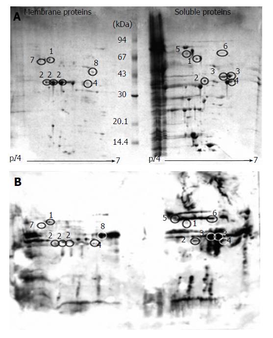
Figure 1 A: Coomassie blue stained 2D gel of S.
flexneri membrane and soluble protein preparations; B: Immunoblot of S. flexneri membrane and soluble protein preparations. Circles indicate immunoreactive spots successfully matched and identified from the Coomassie gel (A). Numbers correspond to Table 1.
- Citation: Jennison AV, Raqib R, Verma NK. Immunoproteome analysis of soluble and membrane proteins of Shigella flexneri 2457T. World J Gastroenterol 2006; 12(41): 6683-6688
- URL: https://www.wjgnet.com/1007-9327/full/v12/i41/6683.htm
- DOI: https://dx.doi.org/10.3748/wjg.v12.i41.6683


