Copyright
©2006 Baishideng Publishing Group Co.
World J Gastroenterol. Oct 14, 2006; 12(38): 6239-6243
Published online Oct 14, 2006. doi: 10.3748/wjg.v12.i38.6239
Published online Oct 14, 2006. doi: 10.3748/wjg.v12.i38.6239
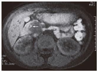
Figure 3 Precontast T1-weighted axial MR image shows that the mass is predominantly hypointense and the hyperintense focus represents hemorrhage (arrow).
- Citation: Karatag O, Yenice G, Ozkurt H, Basak M, Basaran C, Yilmaz B. A case of solid pseudopapillary tumor of the pancreas. World J Gastroenterol 2006; 12(38): 6239-6243
- URL: https://www.wjgnet.com/1007-9327/full/v12/i38/6239.htm
- DOI: https://dx.doi.org/10.3748/wjg.v12.i38.6239


