Copyright
©2006 Baishideng Publishing Group Co.
World J Gastroenterol. Jul 7, 2006; 12(25): 4101-4103
Published online Jul 7, 2006. doi: 10.3748/wjg.v12.i25.4101
Published online Jul 7, 2006. doi: 10.3748/wjg.v12.i25.4101
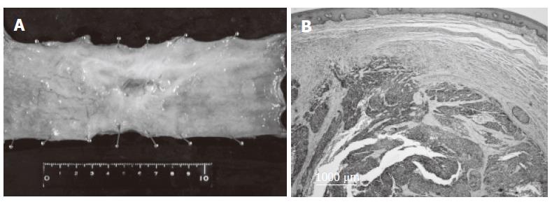
Figure 3 Resected specimen showing a 78 mm × 60 mm superficially invasive tumor (A) and histological examination showing foci of poorly-differentiated squamous cell carcinoma scattered in the submucosal layer (B) (HE, X 20).
- Citation: Kosugi SI, Kanda T, Nishimaki T, Nakagawa S, Yajima K, Ohashi M, Hatakeyama K. Successful treatment for esophageal carcinoma with lung metastasis by induction chemotherapy followed by salvage esophagectomy: Report of a case. World J Gastroenterol 2006; 12(25): 4101-4103
- URL: https://www.wjgnet.com/1007-9327/full/v12/i25/4101.htm
- DOI: https://dx.doi.org/10.3748/wjg.v12.i25.4101


