Copyright
©2006 Baishideng Publishing Group Co.
World J Gastroenterol. Apr 14, 2006; 12(14): 2293-2296
Published online Apr 14, 2006. doi: 10.3748/wjg.v12.i14.2293
Published online Apr 14, 2006. doi: 10.3748/wjg.v12.i14.2293
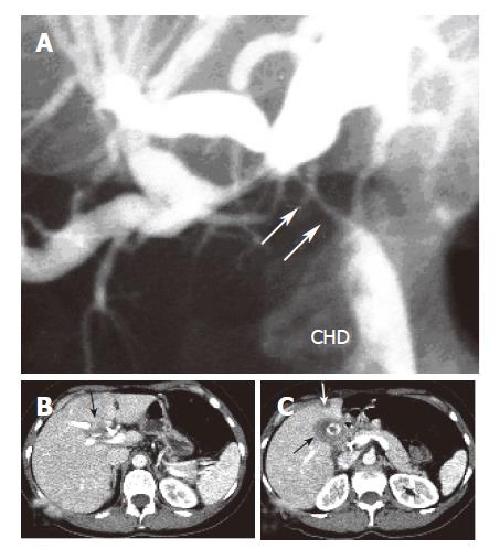
Figure 1 Proximal obstruction (arrows) of the common hepatic bile duct (CHD) with upstream dilatation of both left and right intrahepatic bile ducts (A), a soft tissue mass at the hepatic hilum (B), and thickened gallbladder wall (black arrow) with a concretion and hepatic lesion in liver segment 4 (C).
- Citation: Spinelli A, Schumacher G, Pascher A, Lopez-Hanninen E, Al-Abadi H, Benckert C, Sauer IM, Pratschke J, Neumann UP, Jonas S, Langrehr JM, Neuhaus P. Extended surgical resection for xanthogranulomatous cholecystitis mimicking advanced gallbladder carcinoma: A case report and review of literature. World J Gastroenterol 2006; 12(14): 2293-2296
- URL: https://www.wjgnet.com/1007-9327/full/v12/i14/2293.htm
- DOI: https://dx.doi.org/10.3748/wjg.v12.i14.2293


