Copyright
©2005 Baishideng Publishing Group Inc.
World J Gastroenterol. Feb 14, 2005; 11(6): 922-925
Published online Feb 14, 2005. doi: 10.3748/wjg.v11.i6.922
Published online Feb 14, 2005. doi: 10.3748/wjg.v11.i6.922
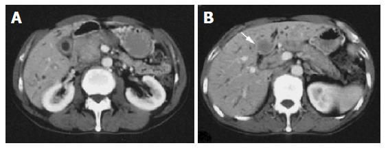
Figure 1 A: CT demonstrated dilatation of the intrahepatic bile ducts, swelling of the pancreatic head, thickening of the gallbladder wall; B: CT revealed the liver mass, which was not enhanced by contrast medium.
- Citation: Sasahira N, Kawabe T, Nakamura A, Shimura K, Shimura H, Itobayashi E, Asada M, Shiratori Y, Omata M. Inflammatory pseudotumor of the liver and peripheral eosinophilia in autoimmune pancreatitis. World J Gastroenterol 2005; 11(6): 922-925
- URL: https://www.wjgnet.com/1007-9327/full/v11/i6/922.htm
- DOI: https://dx.doi.org/10.3748/wjg.v11.i6.922


