Copyright
©2005 Baishideng Publishing Group Inc.
World J Gastroenterol. Feb 7, 2005; 11(5): 686-689
Published online Feb 7, 2005. doi: 10.3748/wjg.v11.i5.686
Published online Feb 7, 2005. doi: 10.3748/wjg.v11.i5.686
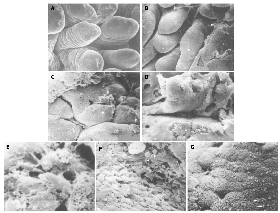
Figure 1 Photomicrograph of scanning electron microscopy of non-specific duodenitis.
A: Villi in uniformed shapes of leaf-like or fingerlike pattern of normal duodenal bulb mucosa. ×200; B: Deformed and integrated villi (asterisk) and exudates (arrow) in degree II of NSD. ×100; C: Broadened, flattened, convoluted or cerebroid villi, masses or band-like mucus (arrows), ulcer-like defect on the surface of a villus (long arrow) in degree II of NSD. ×100; D: Ulcer-like defect (asterisk) and a few of H pylori (arrows) in mucus. ×2500; E: Mucus and severe erosion on the surface of mucosa in degree III of NSD. ×500; F: Macrophages (long arrow) and inflammatory cells (arrows) on the surface of mucosa and gastric metoplasia cells (asterisk). ×2200; G: H pylori in S-shape on the microvilli of epithelial cells (arrows). ×3200.
- Citation: Wang CX, Liu LJ, Guan J, Zhao XL. Ultrastructural changes in non-specific duodenitis. World J Gastroenterol 2005; 11(5): 686-689
- URL: https://www.wjgnet.com/1007-9327/full/v11/i5/686.htm
- DOI: https://dx.doi.org/10.3748/wjg.v11.i5.686


