Copyright
©2005 Baishideng Publishing Group Inc.
World J Gastroenterol. Oct 28, 2005; 11(40): 6348-6353
Published online Oct 28, 2005. doi: 10.3748/wjg.v11.i40.6348
Published online Oct 28, 2005. doi: 10.3748/wjg.v11.i40.6348
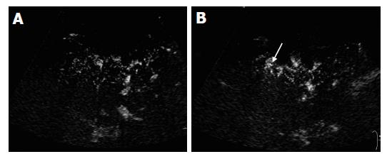
Figure 4 Contrast-enhanced US images in cirrhotic patients.
A and B: On contrast-enhanced US images, the peripheral vessels had poor delineation and were irregular (A). There was no tapering of the vessels. Small branches of the vessels were rarely seen (A), while abnormal ring-like structure (B, arrow) was displayed.
- Citation: Zheng RQ, Zhang B, Kudo M, Sakaguchi Y. Hemodynamic and morphologic changes of peripheral hepatic vasculature in cirrhotic liver disease: A preliminary study using contrast-enhanced coded phase inversion harmonic ultrasonography. World J Gastroenterol 2005; 11(40): 6348-6353
- URL: https://www.wjgnet.com/1007-9327/full/v11/i40/6348.htm
- DOI: https://dx.doi.org/10.3748/wjg.v11.i40.6348


