Copyright
©2005 Baishideng Publishing Group Inc.
World J Gastroenterol. Oct 28, 2005; 11(40): 6348-6353
Published online Oct 28, 2005. doi: 10.3748/wjg.v11.i40.6348
Published online Oct 28, 2005. doi: 10.3748/wjg.v11.i40.6348
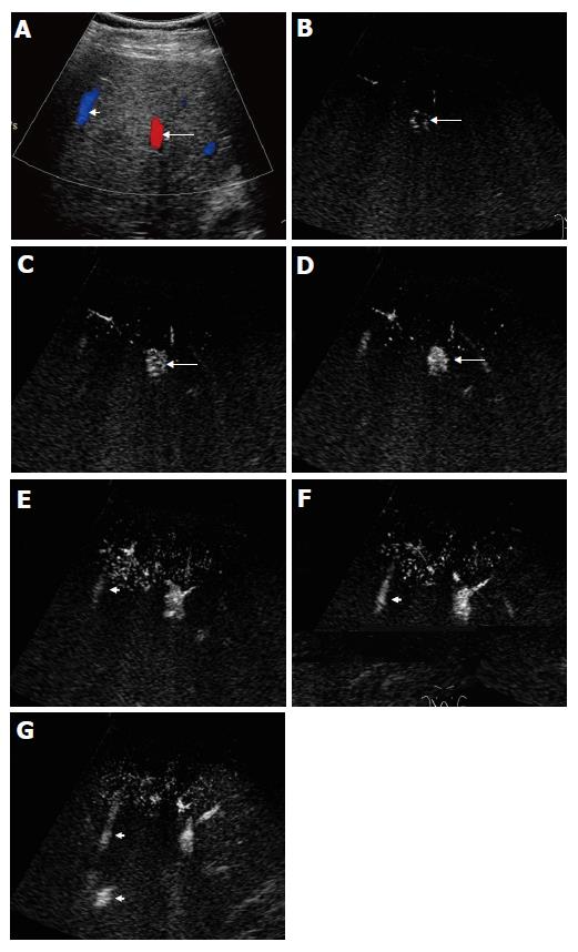
Figure 2 Contrast-enhanced US images in a 32-year-old normal control.
A: Before contrast, color Doppler US was performed to show the location and course of the portal vein (arrow) and hepatic vein (arrowhead); B: In the early arterial phase, the portal vein was not enhanced, while the parallel running artery was displayed by microbubble enhancement (arrow); C: Twenty-eight seconds after injection of Levovist, bright dots of microbubbles began to fill in the portal vein (arrow); D: Four seconds later, the portal vein was obviously enhanced by microbubbles (arrow); E: Before microbubbles arrived, the hepatic vein (arrowhead) was mildly echogenic; F and G: Thirty-seven seconds after injection of contrast medium, the hepatic vein (arrowhead) became more echogenic (F) than it was on E, indicating enhancement by microbubbles, and 4 s later the proximal part of hepatic vein was also clearly seen (arrowheads) (G).
- Citation: Zheng RQ, Zhang B, Kudo M, Sakaguchi Y. Hemodynamic and morphologic changes of peripheral hepatic vasculature in cirrhotic liver disease: A preliminary study using contrast-enhanced coded phase inversion harmonic ultrasonography. World J Gastroenterol 2005; 11(40): 6348-6353
- URL: https://www.wjgnet.com/1007-9327/full/v11/i40/6348.htm
- DOI: https://dx.doi.org/10.3748/wjg.v11.i40.6348


