Copyright
©2005 Baishideng Publishing Group Inc.
World J Gastroenterol. Oct 28, 2005; 11(40): 6338-6347
Published online Oct 28, 2005. doi: 10.3748/wjg.v11.i40.6338
Published online Oct 28, 2005. doi: 10.3748/wjg.v11.i40.6338
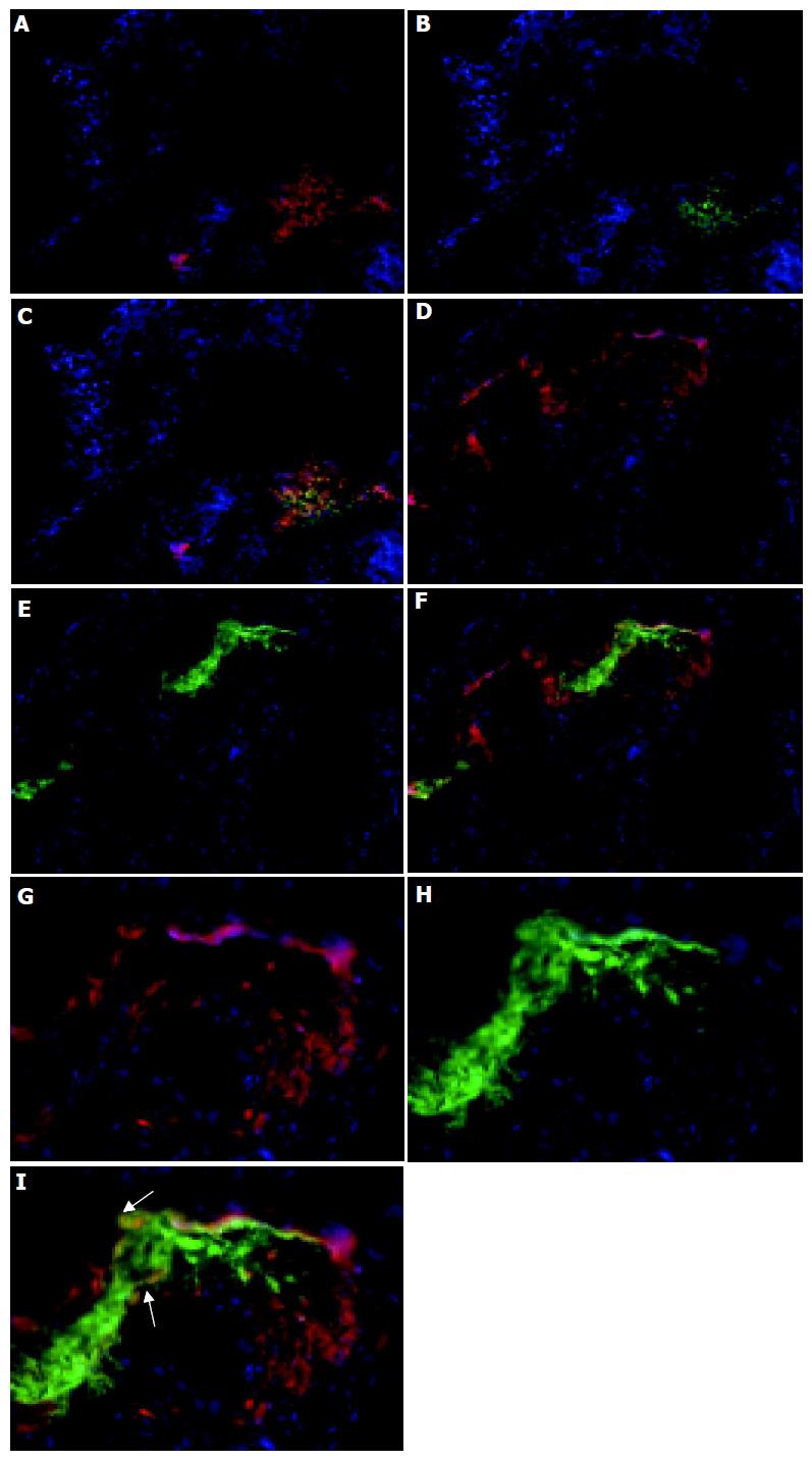
Figure 3 Lymphoid follicles in the colon.
Colon sections were immunolabeled with CD11c and Cy3 to identify DC (red, A, C, D, F, G, and I), CD4 and FITC to identify Th cells (B and C) and B220 and FITC to identify B cells (E, F, H, and I) and counterstained with DAPI. Magnification, ×160. The bottom panel shows that there are B220+ CD11c+ cells (yellow) within the lymphoid follicles of colitic mice (magnification, ×400). The dual stained cells have been arrowed to indicate their localization. Tissue was analyzed from the colons of 5 C57BL/6-IL2-/- and 4 C57BL/6 animals.
- Citation: Cruickshank SM, English NR, Felsburg PJ, Carding SR. Characterization of colonic dendritic cells in normal and colitic mice. World J Gastroenterol 2005; 11(40): 6338-6347
- URL: https://www.wjgnet.com/1007-9327/full/v11/i40/6338.htm
- DOI: https://dx.doi.org/10.3748/wjg.v11.i40.6338


