Copyright
©2005 Baishideng Publishing Group Inc.
World J Gastroenterol. Oct 28, 2005; 11(40): 6338-6347
Published online Oct 28, 2005. doi: 10.3748/wjg.v11.i40.6338
Published online Oct 28, 2005. doi: 10.3748/wjg.v11.i40.6338
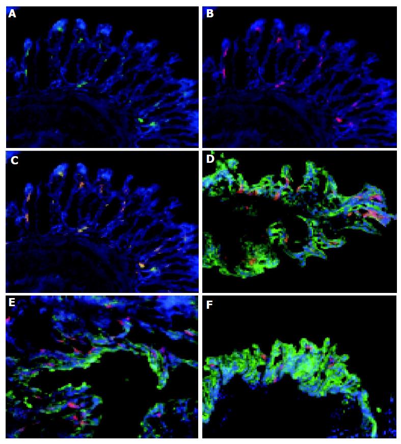
Figure 2 Colonic DC are myeloid DC which are distributed throughout the colon.
Frozen sections of normal colon were stained with MAC-1 and FITC (A), CD11c and Cy3 (B) and counterstained with DAPI. The overlay of the combined images (C) shows that CD11c and MAC-1 co-localized (yellow) consistent with these cells being MAC-1+, CD11c+ myeloid DC. Tissue from the proximal (D), mid (E), and distal (F) colon of normal animals were immunolabeled with CD11c-Cy.3 to identify DC, cytokeratin-FITC to identify epithelial cells and counterstained with DAPI. DC were distributed throughout the colon. Magnification, ×160. Tissue was analyzed from the colons of 5 C57BL/6-IL2-/- and 4 C57BL/6 animals.
- Citation: Cruickshank SM, English NR, Felsburg PJ, Carding SR. Characterization of colonic dendritic cells in normal and colitic mice. World J Gastroenterol 2005; 11(40): 6338-6347
- URL: https://www.wjgnet.com/1007-9327/full/v11/i40/6338.htm
- DOI: https://dx.doi.org/10.3748/wjg.v11.i40.6338


