Copyright
©2005 Baishideng Publishing Group Inc.
World J Gastroenterol. Oct 28, 2005; 11(40): 6338-6347
Published online Oct 28, 2005. doi: 10.3748/wjg.v11.i40.6338
Published online Oct 28, 2005. doi: 10.3748/wjg.v11.i40.6338
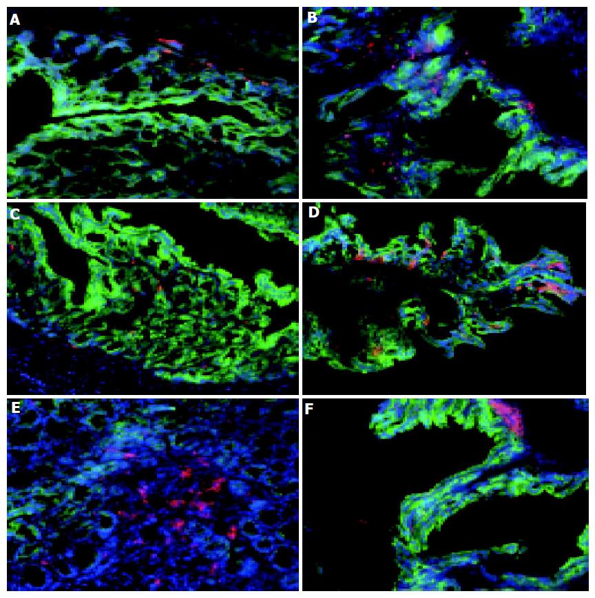
Figure 1 Distribution of macrophages and DC in normal and colitic colon.
Sections of cryopreserved colon from normal C57BL/6 (A, C, and E) and colitic C57BL/6-IL2-/- mice (B, D, and F) were labeled with F4/80 and Cy-3 to identify macrophages (A and B), CD11c and Cy-3 to identify DC (C-F) and cytokeratin-FITC to identify epithelial cells (A-F) and counterstained with DAPI. Although there were increased numbers of macrophages in colitic animals, their distribution was unaltered (A and B). The number of DC was increased in the inflamed colon and their distribution was altered with DC being found in close proximity to the epithelium (C and D). Aggregates of CD11c+ DC were also observed at the base of villi of both normal and colitic colons (E and F). Magnification, ×160. Tissue was analyzed from the colons of 5 C57BL/6-IL2-/- and 4 C57BL/6 animals.
- Citation: Cruickshank SM, English NR, Felsburg PJ, Carding SR. Characterization of colonic dendritic cells in normal and colitic mice. World J Gastroenterol 2005; 11(40): 6338-6347
- URL: https://www.wjgnet.com/1007-9327/full/v11/i40/6338.htm
- DOI: https://dx.doi.org/10.3748/wjg.v11.i40.6338


