Copyright
©The Author(s) 2005.
World J Gastroenterol. Sep 7, 2005; 11(33): 5248-5250
Published online Sep 7, 2005. doi: 10.3748/wjg.v11.i33.5248
Published online Sep 7, 2005. doi: 10.3748/wjg.v11.i33.5248
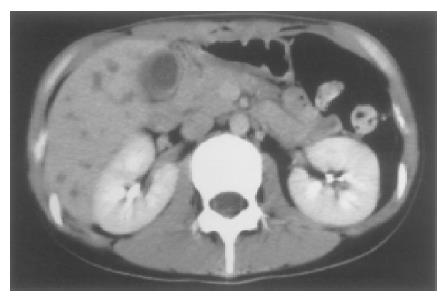
Figure 3 CT showing the gallbladder distended with a thickened wall.
In addition, low density areas faintly enhanced at S6 and S8 in the right lobe of the liver suspected to be abscesses are seen.
- Citation: Suzuki M, Nabeshima K, Miyazaki M, Yoshimura H, Tagawa S, Shiraki K. Churg-Strauss syndrome complicated by colon erosion, acalculous cholecystitis and liver abscesses. World J Gastroenterol 2005; 11(33): 5248-5250
- URL: https://www.wjgnet.com/1007-9327/full/v11/i33/5248.htm
- DOI: https://dx.doi.org/10.3748/wjg.v11.i33.5248


