Copyright
©2005 Baishideng Publishing Group Inc.
World J Gastroenterol. Mar 21, 2005; 11(11): 1719-1721
Published online Mar 21, 2005. doi: 10.3748/wjg.v11.i11.1719
Published online Mar 21, 2005. doi: 10.3748/wjg.v11.i11.1719
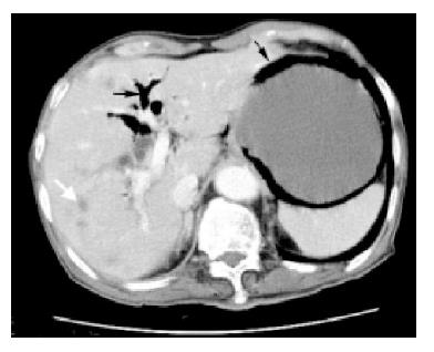
Figure 3 Abdominal computerized tomography revealing multiple liver metastases (white arrow), pneumobilia (thin arrow) and persistent air in the gastric wall (thick arrow).
- Citation: Soon MS, Yen HH, Soon A, Lin OS. Endoscopic ultrasonographic appearance of gastric emphysema. World J Gastroenterol 2005; 11(11): 1719-1721
- URL: https://www.wjgnet.com/1007-9327/full/v11/i11/1719.htm
- DOI: https://dx.doi.org/10.3748/wjg.v11.i11.1719


