Copyright
©The Author(s) 2004.
World J Gastroenterol. Jun 1, 2004; 10(11): 1666-1668
Published online Jun 1, 2004. doi: 10.3748/wjg.v10.i11.1666
Published online Jun 1, 2004. doi: 10.3748/wjg.v10.i11.1666
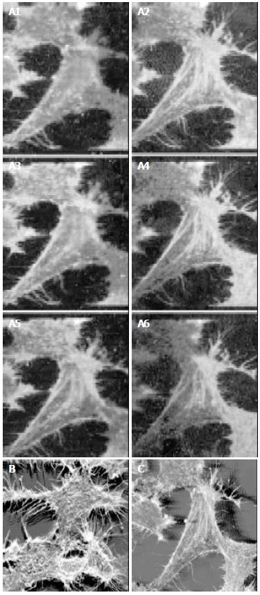
Figure 1 CLSM images of HepG2 cells stained with FITC-phalloidin.
(A: Serial optical sections (1-6) B: Three-dimensional images of Figure 1 serial optical sections. C: Three-dimensional images of HepG2 cells).
- Citation: Huo X, Xu XJ, Chen YW, Yang HW, Piao ZX. Filamentous-actins in human hepatocarcinoma cells with CLSM. World J Gastroenterol 2004; 10(11): 1666-1668
- URL: https://www.wjgnet.com/1007-9327/full/v10/i11/1666.htm
- DOI: https://dx.doi.org/10.3748/wjg.v10.i11.1666


