Copyright
©The Author(s) 2004.
World J Gastroenterol. Jan 1, 2004; 10(1): 152-154
Published online Jan 1, 2004. doi: 10.3748/wjg.v10.i1.152
Published online Jan 1, 2004. doi: 10.3748/wjg.v10.i1.152
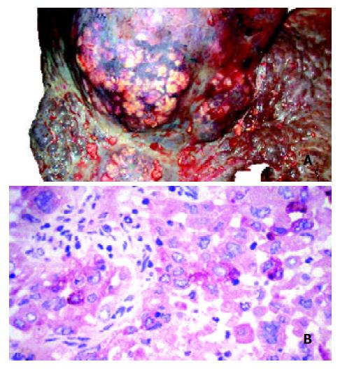
Figure 2 Hepatocellular carcinoma in the presented patient with primary biliary cirrhosis.
A: Gross view of liver at autopsy. Grayish nodules of carcinoma protrude on the surface of the cirrhotic liver. Bar = 1 cm. B: The focal red staining on the histo-logic picture of the hepatocellular carcinoma shows the immu-nohistochemical reaction of AFP. Original magnification 400 ×
- Citation: Horvath A, Folhoffer A, Lakatos PL, Halász J, Illyés G, Schaff Z, Hantos MB, Tekes K, Szalay F. Rising plasma nociceptin level during development of HCC: A case report. World J Gastroenterol 2004; 10(1): 152-154
- URL: https://www.wjgnet.com/1007-9327/full/v10/i1/152.htm
- DOI: https://dx.doi.org/10.3748/wjg.v10.i1.152


