Copyright
©The Author(s) 2001.
World J Gastroenterol. Feb 15, 2001; 7(1): 42-48
Published online Feb 15, 2001. doi: 10.3748/wjg.v7.i1.42
Published online Feb 15, 2001. doi: 10.3748/wjg.v7.i1.42
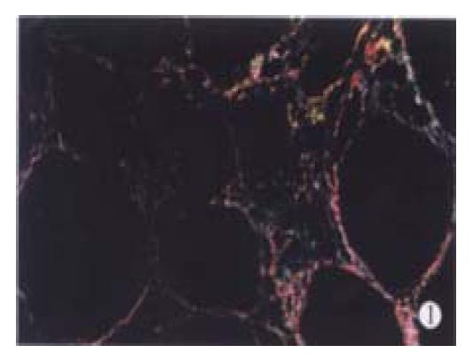
Figure 1 Histological section of rat liver after 12-weeks of CCl4 treatment under polarizes after staining with Sirius Red.
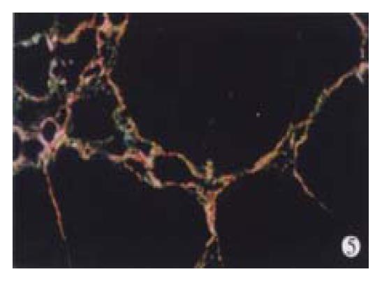
Red and yellow represented collagen type I and green collagen type III (the same below). The pattern of micronodular cirrhosis is evident. × 100
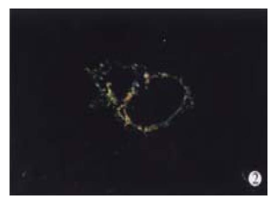
Figure 2 Histological section of rat liver after 8-weeks of treatment with high-dose IFN-γ and 12 weeks of CCl4 under polarizes after staining with Sirius Red.
× 100
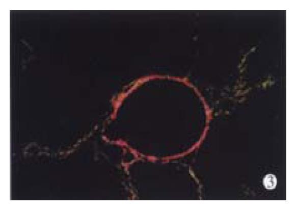
Figure 3 Histological section of rat liver after 8-weeks of treatment with medium-dose IFN-γ and 12 weeks of CCl4 under polarizes after staining with Sirius Red.
× 100
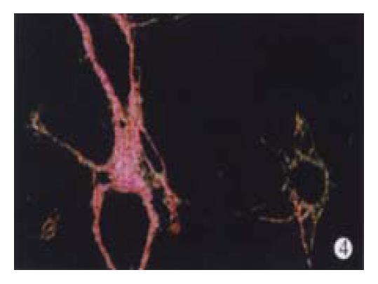
Figure 4 Histological section of rat liver after 8-weeks of treatment with low-dose IFN-γ and 12 weeks of CCl4 under polarizes after staining with Sirius Red.
× 100
Figure 5 Histological section of rat liver after 8-weeks of treatment with colchicine and 12 weeks of CCl4 under polarizes after staining with Sirius Red.
× 100
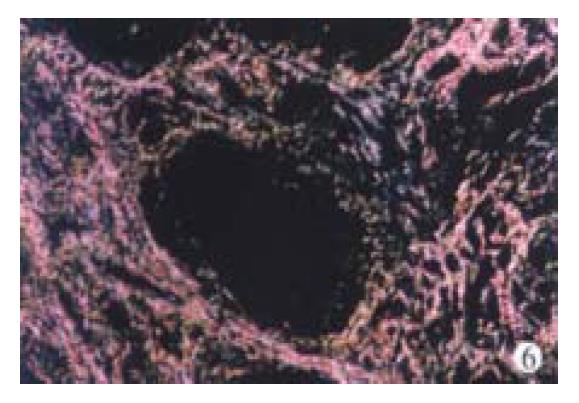
Figure 6 Histological section of liver biopsy of a patient with chronic hepatitis B before IFN-γ treatment under polarizes after staining with Sirius Red.
Figure 6 show two different fields. × 100
- Citation: Weng HL, Cai WM, Liu RH. Animal experiment and clinical study of effect of gamma-interferon on hepatic fibrosis. World J Gastroenterol 2001; 7(1): 42-48
- URL: https://www.wjgnet.com/1007-9327/full/v7/i1/42.htm
- DOI: https://dx.doi.org/10.3748/wjg.v7.i1.42


