Copyright
©The Author(s) 1997.
World J Gastroenterol. Jun 15, 1997; 3(2): 95-97
Published online Jun 15, 1997. doi: 10.3748/wjg.v3.i2.95
Published online Jun 15, 1997. doi: 10.3748/wjg.v3.i2.95
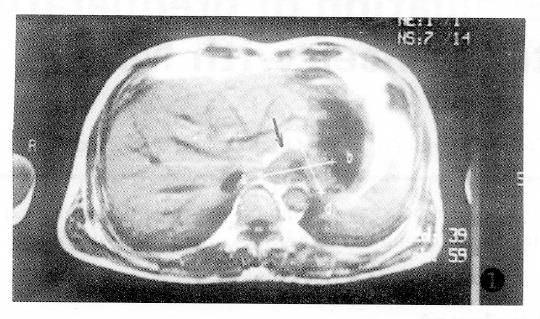
Figure 1 Grade MRT2 tumor, T1WI.
Axial images show a smooth low signal intensity band around the lesion (↑).
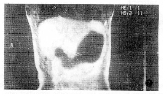
Figure 2 Grade MRT2 tumor, T1WI.
Coronal images show a smooth low signal intensity band around the lesion (↑).
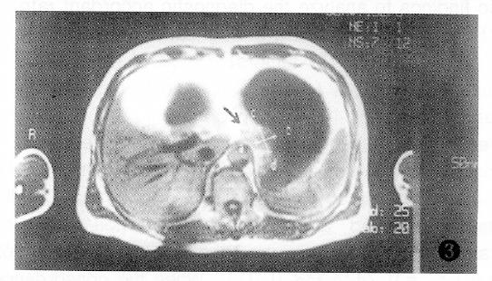
Figure 3 Grade MRT3 tumor, T1WI.
Axial image shows an obscure hypointense band and blurring fat plane outside of the tumor (↑).
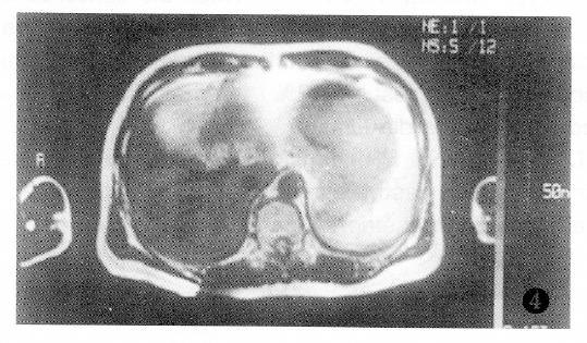
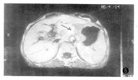
Figure 5 Grade MRT4 tumor, T1WI.
Fat interval between stomach and hepatic left lobe disappeared and was occupied by a isointense tumor.
- Citation: Tang GY, Guo QL, Xin PP. Evaluation of preoperative staging in advanced gastric cancer with MRI. World J Gastroenterol 1997; 3(2): 95-97
- URL: https://www.wjgnet.com/1007-9327/full/v3/i2/95.htm
- DOI: https://dx.doi.org/10.3748/wjg.v3.i2.95


